 Movie
Movie Controller


+ Open data

- Basic information
| Entry | Database: PDB / ID: 6k2e | ||||||
|---|---|---|---|---|---|---|---|
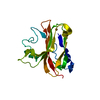
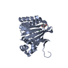
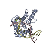
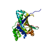
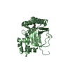
| Title | Crystal structure of cas2 | ||||||
 Components Components | CRISPR/cas2 protein | ||||||
 Keywords Keywords | RNA BINDING PROTEIN / crispr / cas2 | ||||||
| Function / homology | Alpha-Beta Plaits - #240 / Alpha-Beta Plaits / 2-Layer Sandwich / Alpha Beta Function and homology information Function and homology information | ||||||
| Biological species |   Pyrococcus horikoshii OT3 (archaea) Pyrococcus horikoshii OT3 (archaea) | ||||||
| Method |  X-RAY DIFFRACTION / MOLECULAR REPLACEMENT / Resolution: 2.8 Å X-RAY DIFFRACTION / MOLECULAR REPLACEMENT / Resolution: 2.8 Å | ||||||
 Authors Authors | Mo, X. / Bi, M. / Yuan, A.Y. | ||||||
 Citation Citation | Journal: To be published Title: Structural insight into endonuclease CRISPR-associated Cas2 protein Authors: Mo, X. | ||||||
| History |
|
- Structure visualization
| Structure viewer | Molecule: MolmilJmol/JSmol |
|---|
- Downloads & links
-Download
| PDBx/mmCIF format | 6k2e.cif.gz | 48.5 KB | Display | PDBx/mmCIF format |
|---|---|---|---|---|
| PDB format | pdb6k2e.ent.gz | 33.9 KB | Display | PDB format |
| PDBx/mmJSON format | 6k2e.json.gz | Tree view | PDBx/mmJSON format | |
| Others |  Other downloads Other downloads |
-Validation report
| Arichive directory | https://data.pdbj.org/pub/pdb/validation_reports/k2/6k2eftp://data.pdbj.org/pub/pdb/validation_reports/k2/6k2e | HTTPS FTP |
|---|
-Related structure data
| Related structure data | 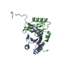 6jhzC  5g4dS C: citing same article ( S: Starting model for refinement |
|---|---|
| Similar structure data |

-Links
 PDBj
PDBj
- Assembly
| Deposited unit | 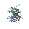
| ||||||||
|---|---|---|---|---|---|---|---|---|---|
| 1 |
| ||||||||
| Unit cell |
|
-Components
| #1: Protein | Mass: 10472.217 Da / Num. of mol.: 2 Source method: isolated from a genetically manipulated source Source: (gene. exp.) Pyrococcus horikoshii OT3 (archaea) / Production host:  #2: Water | ChemComp-HOH / |  Mass: 18.015 Da / Num. of mol.: 1 / Source method: isolated from a natural source / Formula: H2O Mass: 18.015 Da / Num. of mol.: 1 / Source method: isolated from a natural source / Formula: H2O |
|---|
-Experimental details
-Experiment
| Experiment | Method: X-RAY DIFFRACTION / Number of used crystals: 1 |
|---|
- Sample preparation
| Crystal | Density Matthews: 2.8 Å3/Da / Density % sol: 56.07 % |
|---|---|
| Crystal grow | Temperature: 280 K / Method: vapor diffusion, hanging drop Details: 0.02M Magnesium, 16.5% v/v 2-propanol, 0.1M Sodium Cacodylate pH 6.6 |
-Data collection
| Diffraction | Mean temperature: 100 K / Serial crystal experiment: N |
|---|---|
| Diffraction source | Source: ROTATING ANODE / Type: RIGAKU / Wavelength: 1.54 Å |
| Detector | Type: RIGAKU RAXIS II / Detector: IMAGE PLATE / Date: May 14, 2019 |
| Radiation | Protocol: SINGLE WAVELENGTH / Monochromatic (M) / Laue (L): M / Scattering type: x-ray |
| Radiation wavelength | Wavelength: 1.54 Å / Relative weight: 1 |
| Reflection | Resolution: 2.8→25.42 Å / Num. obs: 5885 / % possible obs: 99.73 % / Redundancy: 8 % / Net I/σ(I): 12 |
| Reflection shell | Resolution: 2.8→2.9 Å / Num. unique obs: 587 |
- Processing
| Software |
| |||||||||||||||||||||||||||||||||||
|---|---|---|---|---|---|---|---|---|---|---|---|---|---|---|---|---|---|---|---|---|---|---|---|---|---|---|---|---|---|---|---|---|---|---|---|---|
| Refinement | Method to determine structure: MOLECULAR REPLACEMENT Starting model: 5G4D Resolution: 2.8→25.42 Å / SU ML: 0.47 / Cross valid method: FREE R-VALUE / σ(F): 1.35 / Phase error: 28.03
| |||||||||||||||||||||||||||||||||||
| Solvent computation | Shrinkage radii: 0.9 Å / VDW probe radii: 1.11 Å | |||||||||||||||||||||||||||||||||||
| Refinement step | Cycle: LAST / Resolution: 2.8→25.42 Å
| |||||||||||||||||||||||||||||||||||
| Refine LS restraints |
| |||||||||||||||||||||||||||||||||||
| LS refinement shell |
|
