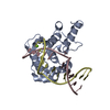
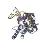
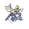
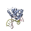
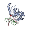
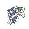
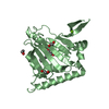
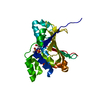
satellite DNA binding / pyrimidine-specific mismatch base pair DNA N-glycosylase activity / depyrimidination / DNA N-glycosylase activity / Displacement of DNA glycosylase by APEX1 / Hydrolases; Glycosylases; Hydrolysing N-glycosyl compounds / Recognition and association of DNA glycosylase with site containing an affected pyrimidine / Cleavage of the damaged pyrimidine / DNA endonuclease activity / response to estradiol ...satellite DNA binding / pyrimidine-specific mismatch base pair DNA N-glycosylase activity / depyrimidination / DNA N-glycosylase activity / Displacement of DNA glycosylase by APEX1 / Hydrolases; Glycosylases; Hydrolysing N-glycosyl compounds / Recognition and association of DNA glycosylase with site containing an affected pyrimidine / Cleavage of the damaged pyrimidine / DNA endonuclease activity / response to estradiol / nuclear speck / DNA repair / DNA binding / nucleoplasm / nucleus Similarity search - Function
Resolution: 1.83→38.91 Å / Cor.coef. Fo:Fc: 0.961 / Cor.coef. Fo:Fc free: 0.947 / SU B: 2.48 / SU ML: 0.077 / Cross valid method: THROUGHOUT / ESU R: 0.128 / ESU R Free: 0.121 / Stereochemistry target values: MAXIMUM LIKELIHOOD / Details: HYDROGENS HAVE BEEN ADDED IN THE RIDING POSITIONS
Rfactor
Num. reflection
% reflection
Selection details
Rfree
0.20899
1067
5.1 %
RANDOM
Rwork
0.17369
-
-
-
obs
0.17546
19653
94.28 %
-
Solvent computation
Ion probe radii: 0.8 Å / Shrinkage radii: 0.8 Å / VDW probe radii: 1.2 Å / Solvent model: MASK
 Movie
Movie Controller


 Open data
 Basic information
 Components
 Keywords
 Function and homology information
 Homo sapiens (human)
 X-RAY DIFFRACTION /
  Authors
 United States, 2items
  Citation
 Structure visualization
 Downloads & links
 Other downloads


 PDBj
PDBj


 Assembly


 Mass: 24.305 Da / Num. of mol.: 1 / Source method: obtained synthetically / Formula: Mg
 Sample preparation
 Processing