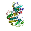
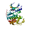
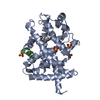
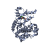
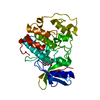
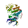
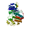
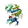
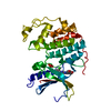
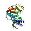
[English] 日本語
Yorodumi
- PDB-6jzd: Crystal structure of peptide-bound VASH2-SVBP complex -

+
Open data


ID or keywords:

Loading...

-
Basic information

Entry
Database: PDB / ID: 6jzd
TitleCrystal structure of peptide-bound VASH2-SVBP complex
Components
  • GLU-GLY-GLU-GLU-TYR
  • Small vasohibin-binding protein
  • Tubulinyl-Tyr carboxypeptidase 2
KeywordsCYTOSOLIC PROTEIN / VASH2 / SVBP / tubulin detyrosination
Function / homology
Function and homology information


cell-cell fusion / Microtubule-dependent trafficking of connexons from Golgi to the plasma membrane / Cilium Assembly / Sealing of the nuclear envelope (NE) by ESCRT-III / regulation of metallopeptidase activity / syncytium formation by plasma membrane fusion / Intraflagellar transport / tubulinyl-Tyr carboxypeptidase / tubulin-tyrosine carboxypeptidase / Carboxyterminal post-translational modifications of tubulin ...cell-cell fusion / Microtubule-dependent trafficking of connexons from Golgi to the plasma membrane / Cilium Assembly / Sealing of the nuclear envelope (NE) by ESCRT-III / regulation of metallopeptidase activity / syncytium formation by plasma membrane fusion / Intraflagellar transport / tubulinyl-Tyr carboxypeptidase / tubulin-tyrosine carboxypeptidase / Carboxyterminal post-translational modifications of tubulin / COPI-independent Golgi-to-ER retrograde traffic / HSP90 chaperone cycle for steroid hormone receptors (SHR) in the presence of ligand / PKR-mediated signaling / COPI-mediated anterograde transport / Aggrephagy / Kinesins / Mitotic Prometaphase / EML4 and NUDC in mitotic spindle formation / Resolution of Sister Chromatid Cohesion / RHO GTPases activate IQGAPs / The role of GTSE1 in G2/M progression after G2 checkpoint / Recycling pathway of L1 / axonemal microtubule / COPI-dependent Golgi-to-ER retrograde traffic / RHO GTPases Activate Formins / Carboxyterminal post-translational modifications of tubulin / Separation of Sister Chromatids / Hedgehog 'off' state / organelle transport along microtubule / glial cell differentiation / Loss of Nlp from mitotic centrosomes / Recruitment of mitotic centrosome proteins and complexes / Loss of proteins required for interphase microtubule organization from the centrosome / Recruitment of NuMA to mitotic centrosomes / Anchoring of the basal body to the plasma membrane / peptidase activator activity / AURKA Activation by TPX2 / forebrain morphogenesis / placenta blood vessel development / neuron projection arborization / Regulation of PLK1 Activity at G2/M Transition / labyrinthine layer development / cerebellar cortex morphogenesis / dentate gyrus development / MHC class II antigen presentation / pyramidal neuron differentiation / centrosome cycle / motor behavior / labyrinthine layer blood vessel development / negative regulation of endothelial cell migration / response to L-glutamate / smoothened signaling pathway / adult behavior / regulation of synapse organization / axon development / startle response / locomotory exploration behavior / microtubule polymerization / cytoplasmic microtubule / protein secretion / response to tumor necrosis factor / regulation of angiogenesis / metallocarboxypeptidase activity / response to mechanical stimulus / condensed chromosome / homeostasis of number of cells within a tissue / negative regulation of protein ubiquitination / positive regulation of endothelial cell proliferation / cellular response to calcium ion / adult locomotory behavior / neurogenesis / locomotory behavior / hippocampus development / Hydrolases; Acting on acid anhydrides; Acting on GTP to facilitate cellular and subcellular movement / synapse organization / intracellular protein transport / neuron migration / visual learning / neuromuscular junction / neuron differentiation / recycling endosome / structural constituent of cytoskeleton / cerebral cortex development / memory / cytoplasmic ribonucleoprotein granule / microtubule cytoskeleton organization / positive regulation of angiogenesis / apical part of cell / myelin sheath / actin binding / mitotic cell cycle / gene expression / microtubule binding / neuron apoptotic process / microtubule / cytoskeleton / hydrolase activity / membrane raft / protein heterodimerization activity / protein domain specific binding
Similarity search - Function
Tubulinyl-Tyr carboxypeptidase / Small vasohibin-binding protein / Vasohibin / Coiled-coil domain-containing protein 23 / Alpha tubulin / Tubulin / Tubulin, C-terminal / Tubulin C-terminal domain / Tubulin, conserved site / Tubulin subunits alpha, beta, and gamma signature. ...Tubulinyl-Tyr carboxypeptidase / Small vasohibin-binding protein / Vasohibin / Coiled-coil domain-containing protein 23 / Alpha tubulin / Tubulin / Tubulin, C-terminal / Tubulin C-terminal domain / Tubulin, conserved site / Tubulin subunits alpha, beta, and gamma signature. / Tubulin/FtsZ family, C-terminal domain / Tubulin/FtsZ-like, C-terminal domain / Tubulin/FtsZ, C-terminal / Tubulin/FtsZ, 2-layer sandwich domain / Tubulin/FtsZ family, GTPase domain / Tubulin/FtsZ family, GTPase domain / Tubulin/FtsZ, GTPase domain / Tubulin/FtsZ, GTPase domain superfamily
Similarity search - Domain/homology
Tubulin alpha-1A chain / Tubulinyl-Tyr carboxypeptidase 2 / Small vasohibin-binding protein
Similarity search - Component
Biological speciesMus musculus (house mouse)
Homo sapiens (human)
MethodX-RAY DIFFRACTION / SYNCHROTRON / MOLECULAR REPLACEMENT / Resolution: 2.479 Å
AuthorsChen, Z. / Ling, Y. / Zeyuan, G. / Zhu, L.
CitationJournal: Nat Commun / Year: 2019
Title: Structural basis of tubulin detyrosination by VASH2/SVBP heterodimer.
Authors: Zhou, C. / Yan, L. / Zhang, W.H. / Liu, Z.
History
DepositionMay 1, 2019Deposition site: PDBJ / Processing site: PDBJ
Revision 1.0Jul 17, 2019Provider: repository / Type: Initial release
Revision 1.1Jul 24, 2019Group: Data collection / Database references / Category: citation / citation_author / Item: _citation.title / _citation_author.name
Revision 1.2Aug 7, 2019Group: Data collection / Database references / Category: citation / citation_author
Item: _citation.journal_volume / _citation.page_first ..._citation.journal_volume / _citation.page_first / _citation.page_last / _citation.pdbx_database_id_PubMed / _citation_author.name
Revision 1.3Mar 27, 2024Group: Data collection / Database references / Category: chem_comp_atom / chem_comp_bond / database_2
Item: _database_2.pdbx_DOI / _database_2.pdbx_database_accession

-
Structure visualization

Structure viewerMolecule:
MolmilJmol/JSmol

Downloads & links

-
Assembly

Deposited unit
A: Tubulinyl-Tyr carboxypeptidase 2
B: Small vasohibin-binding protein
C: GLU-GLY-GLU-GLU-TYR


Theoretical massNumber of molelcules
Total (without water)52,5413
Polymers52,5413
Non-polymers00
Water28816
1


  • Idetical with deposited unit
  • defined by author&software
  • Evidence: gel filtration
TypeNameSymmetry operationNumber
identity operation1_555x,y,z1
Buried area3710 Å2
ΔGint-21 kcal/mol
Surface area14500 Å2
MethodPISA
Unit cell
Length a, b, c (Å)74.004, 111.339, 126.317
Angle α, β, γ (deg.)90.00, 90.00, 90.00
Int Tables number20
Space group name H-MC2221

-
Components

#1: Protein Tubulinyl-Tyr carboxypeptidase 2 / Vasohibin-2 / Vasohibin-like protein


Mass: 40536.906 Da / Num. of mol.: 1 / Mutation: C158A
Source method: isolated from a genetically manipulated source
Source: (gene. exp.) Mus musculus (house mouse) / Gene: Vash2, Vashl / Production host: 
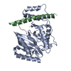
Spodoptera frugiperda (fall armyworm) / References: UniProt: Q8C5G2, tubulinyl-Tyr carboxypeptidase
#2: Protein Small vasohibin-binding protein / Coiled coil domain-containing protein 23


Mass: 10347.556 Da / Num. of mol.: 1
Source method: isolated from a genetically manipulated source
Source: (gene. exp.) Homo sapiens (human) / Gene: SVBP, CCDC23 / Production host: Spodoptera frugiperda (fall armyworm) / References: UniProt: Q8N300
#3: Protein/peptide GLU-GLY-GLU-GLU-TYR


Mass: 1656.568 Da / Num. of mol.: 1 / Source method: obtained synthetically / Source: (synth.) Mus musculus (house mouse) / References: UniProt: P68369*PLUS
#4: Water ChemComp-HOH / water


Mass: 18.015 Da / Num. of mol.: 16 / Source method: isolated from a natural source / Formula: H2O

-
Experimental details

-
Experiment

ExperimentMethod: X-RAY DIFFRACTION / Number of used crystals: 1

-
Sample preparation

CrystalDensity Matthews: 2.48 Å3/Da / Density % sol: 50.33 %
Crystal growTemperature: 291.15 K / Method: vapor diffusion, hanging drop / pH: 7 / Details: HEPES, potassium chloride, Ammonium sulphate

-
Data collection

DiffractionMean temperature: 100 K / Serial crystal experiment: N
Diffraction sourceSource: SYNCHROTRON / Site: SSRF / Beamline: BL17U1 / Wavelength: 0.9791 Å
DetectorType: DECTRIS EIGER X 16M / Detector: PIXEL / Date: Jan 5, 2019
RadiationProtocol: SINGLE WAVELENGTH / Monochromatic (M) / Laue (L): M / Scattering type: x-ray
Radiation wavelengthWavelength: 0.9791 Å / Relative weight: 1
ReflectionResolution: 2.45→50 Å / Num. obs: 18801 / % possible obs: 99.6 % / Redundancy: 6.4 % / CC1/2: 0.999 / Rmerge(I) obs: 0.059 / Rpim(I) all: 0.025 / Net I/σ(I): 17.1
Reflection shellResolution: 2.48→2.58 Å / Redundancy: 6.1 % / Rmerge(I) obs: 0.759 / Num. unique obs: 2048 / CC1/2: 0.764 / Rpim(I) all: 0.332 / % possible all: 98.5

-
Processing

Software
NameVersionClassification
PHENIX(1.14rc3_3206: ???)refinement
XDSdata reduction
Aimlessdata scaling
PHASERphasing
RefinementMethod to determine structure: MOLECULAR REPLACEMENT / Resolution: 2.479→44.11 Å / SU ML: 0.32 / Cross valid method: FREE R-VALUE / σ(F): 1.33 / Phase error: 23.19 / Stereochemistry target values: ML
RfactorNum. reflection% reflection
Rfree0.2296 911 4.85 %
Rwork0.1793 --
obs0.1816 18768 99.43 %
Solvent computationShrinkage radii: 0.9 Å / VDW probe radii: 1.11 Å / Solvent model: FLAT BULK SOLVENT MODEL
Refinement stepCycle: LAST / Resolution: 2.479→44.11 Å
ProteinNucleic acidLigandSolventTotal
Num. atoms2402 0 0 16 2418
Refine LS restraints
Refine-IDTypeDev idealNumber
X-RAY DIFFRACTIONf_bond_d0.0082462
X-RAY DIFFRACTIONf_angle_d0.9433315
X-RAY DIFFRACTIONf_dihedral_angle_d16.1041492
X-RAY DIFFRACTIONf_chiral_restr0.053348
X-RAY DIFFRACTIONf_plane_restr0.005422
LS refinement shell
Resolution (Å)Rfactor RfreeNum. reflection RfreeRfactor RworkNum. reflection RworkRefine-ID% reflection obs (%)
2.4787-2.60940.31891380.25112448X-RAY DIFFRACTION98
2.6094-2.77280.25391390.22912528X-RAY DIFFRACTION100
2.7728-2.98690.27831250.20942530X-RAY DIFFRACTION100
2.9869-3.28740.27591300.21442541X-RAY DIFFRACTION100
3.2874-3.76290.26351310.19022559X-RAY DIFFRACTION99
3.7629-4.73990.19531160.15332584X-RAY DIFFRACTION99
4.7399-44.11710.19661320.16462667X-RAY DIFFRACTION99
Refinement TLS params.Method: refined / Origin x: 3.8197 Å / Origin y: 22.1721 Å / Origin z: -9.8623 Å
111213212223313233
T0.4035 Å2-0.0288 Å20.022 Å2-0.4845 Å2-0.0196 Å2--0.4206 Å2
L2.0739 °2-0.3446 °2-0.5696 °2-3.02 °20.3909 °2--2.0512 °2
S0.0733 Å °0.0034 Å °0.2797 Å °-0.1396 Å °0.0885 Å °-0.0598 Å °-0.183 Å °0.0303 Å °-0.1645 Å °
Refinement TLS groupSelection details: all

+
About Yorodumi

-
News

-
Feb 9, 2022. New format data for meta-information of EMDB entries

New format data for meta-information of EMDB entries

  • Version 3 of the EMDB header file is now the official format.
  • The previous official version 1.9 will be removed from the archive.

Related info.:EMDB header

External links:wwPDB to switch to version 3 of the EMDB data model

-
Aug 12, 2020. Covid-19 info

Covid-19 info

URL: https://pdbjlvh1.pdbj.org/emnavi/covid19.php

New page: Covid-19 featured information page in EM Navigator.

Related info.:Covid-19 info / Mar 5, 2020. Novel coronavirus structure data

+
Mar 5, 2020. Novel coronavirus structure data

Novel coronavirus structure data

Related info.:Yorodumi Speices / Aug 12, 2020. Covid-19 info

External links:COVID-19 featured content - PDBj / Molecule of the Month (242):Coronavirus Proteases

+
Jan 31, 2019. EMDB accession codes are about to change! (news from PDBe EMDB page)

EMDB accession codes are about to change! (news from PDBe EMDB page)

  • The allocation of 4 digits for EMDB accession codes will soon come to an end. Whilst these codes will remain in use, new EMDB accession codes will include an additional digit and will expand incrementally as the available range of codes is exhausted. The current 4-digit format prefixed with “EMD-” (i.e. EMD-XXXX) will advance to a 5-digit format (i.e. EMD-XXXXX), and so on. It is currently estimated that the 4-digit codes will be depleted around Spring 2019, at which point the 5-digit format will come into force.
  • The EM Navigator/Yorodumi systems omit the EMD- prefix.

Related info.:Q: What is EMD? / ID/Accession-code notation in Yorodumi/EM Navigator

External links:EMDB Accession Codes are Changing Soon! / Contact to PDBj

+
Jul 12, 2017. Major update of PDB

Major update of PDB

  • wwPDB released updated PDB data conforming to the new PDBx/mmCIF dictionary.
  • This is a major update changing the version number from 4 to 5, and with Remediation, in which all the entries are updated.
  • In this update, many items about electron microscopy experimental information are reorganized (e.g. em_software).
  • Now, EM Navigator and Yorodumi are based on the updated data.

External links:wwPDB Remediation / Enriched Model Files Conforming to OneDep Data Standards Now Available in the PDB FTP Archive

-
Yorodumi

Thousand views of thousand structures

  • Yorodumi is a browser for structure data from EMDB, PDB, SASBDB, etc.
  • This page is also the successor to EM Navigator detail page, and also detail information page/front-end page for Omokage search.
  • The word "yorodu" (or yorozu) is an old Japanese word meaning "ten thousand". "mi" (miru) is to see.

Related info.:EMDB / PDB / SASBDB / Comparison of 3 databanks / Yorodumi Search / Aug 31, 2016. New EM Navigator & Yorodumi / Yorodumi Papers / Jmol/JSmol / Function and homology information / Changes in new EM Navigator and Yorodumi

Read more