 Movie
Movie Controller

[English] 日本語
 Yorodumi
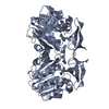
- PDB-6jqw: Crystal structure of a hydrogenase from Trichosporon moniliiforme -
+ Open data

- Basic information
| Entry | Database: PDB / ID: 6jqw | ||||||
|---|---|---|---|---|---|---|---|
| Title | Crystal structure of a hydrogenase from Trichosporon moniliiforme | ||||||
 Components Components | Salicylate decarboxylase | ||||||
 Keywords Keywords | HYDROLASE / Structure function / Salicylate / decarboxylase | ||||||
| Function / homology |  Function and homology information Function and homology informationsalicylate decarboxylase / secondary metabolic process / carboxy-lyase activity / hydrolase activity / cytosol Similarity search - Function | ||||||
| Biological species |  Cutaneotrichosporon moniliiforme (fungus) Cutaneotrichosporon moniliiforme (fungus) | ||||||
| Method |  X-RAY DIFFRACTION / SYNCHROTRON / MOLECULAR REPLACEMENT / molecular replacement / Resolution: 1.437 Å X-RAY DIFFRACTION / SYNCHROTRON / MOLECULAR REPLACEMENT / molecular replacement / Resolution: 1.437 Å | ||||||
 Authors Authors | Qin, H.M. / Chen, X.T. | ||||||
 Citation Citation | Journal: J.Agric.Food Chem. / Year: 2021 Title: Structural Basis of Salicylic Acid Decarboxylase Reveals a Unique Substrate Recognition Mode and Access Channel. Authors: Gao, X. / Wu, M. / Zhang, W. / Li, C. / Guo, R.T. / Dai, Y. / Liu, W. / Mao, S. / Lu, F. / Qin, H.M. | ||||||
| History |
|
- Structure visualization
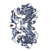
| Structure viewer | Molecule: MolmilJmol/JSmol |
|---|
- Downloads & links
-Download
| PDBx/mmCIF format | 6jqw.cif.gz | 314.6 KB | Display | PDBx/mmCIF format |
|---|---|---|---|---|
| PDB format | pdb6jqw.ent.gz | 253.8 KB | Display | PDB format |
| PDBx/mmJSON format | 6jqw.json.gz | Tree view | PDBx/mmJSON format | |
| Others |  Other downloads Other downloads |
-Validation report
| Summary document | 6jqw_validation.pdf.gz | 432.5 KB | Display | wwPDB validaton report |
|---|---|---|---|---|
| Full document | 6jqw_full_validation.pdf.gz | 434.3 KB | Display | |
| Data in XML | 6jqw_validation.xml.gz | 32.8 KB | Display | |
| Data in CIF | 6jqw_validation.cif.gz | 51.5 KB | Display | |
| Arichive directory | https://data.pdbj.org/pub/pdb/validation_reports/jq/6jqwftp://data.pdbj.org/pub/pdb/validation_reports/jq/6jqw | HTTPS FTP |
-Related structure data
| Related structure data |  6jqxC  2dvtS S: Starting model for refinement C: citing same article ( |
|---|---|
| Similar structure data |


-Links
 PDBj
PDBj

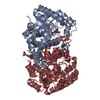
- Assembly
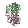
| Deposited unit | 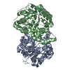
| ||||||||
|---|---|---|---|---|---|---|---|---|---|
| 1 |
| ||||||||
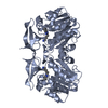
| Unit cell |
|
-Components
| #1: Protein | Mass: 40157.145 Da / Num. of mol.: 2 Source method: isolated from a genetically manipulated source Source: (gene. exp.) Cutaneotrichosporon moniliiforme (fungus)Gene: sdc / Plasmid: pQE-80L / Production host:  #2: Chemical |   Mass: 65.409 Da / Num. of mol.: 2 / Source method: obtained synthetically / Formula: Zn Mass: 65.409 Da / Num. of mol.: 2 / Source method: obtained synthetically / Formula: Zn#3: Water | ChemComp-HOH / |  Mass: 18.015 Da / Num. of mol.: 829 / Source method: isolated from a natural source / Formula: H2O Mass: 18.015 Da / Num. of mol.: 829 / Source method: isolated from a natural source / Formula: H2O |
|---|
-Experimental details
-Experiment
| Experiment | Method: X-RAY DIFFRACTION / Number of used crystals: 1 |
|---|
- Sample preparation
| Crystal | Density Matthews: 2.17 Å3/Da / Density % sol: 43.29 % / Mosaicity: 0.603 ° |
|---|---|
| Crystal grow | Temperature: 295 K / Method: vapor diffusion, sitting drop / pH: 8.5 / Details: PEG400, NaCl |
-Data collection
| Diffraction | Mean temperature: 100 K / Serial crystal experiment: N | |||||||||||||||||||||||||||||||||||||||||||||||||||||||||||||||||||||||||||||||||||||||||||||||||||
|---|---|---|---|---|---|---|---|---|---|---|---|---|---|---|---|---|---|---|---|---|---|---|---|---|---|---|---|---|---|---|---|---|---|---|---|---|---|---|---|---|---|---|---|---|---|---|---|---|---|---|---|---|---|---|---|---|---|---|---|---|---|---|---|---|---|---|---|---|---|---|---|---|---|---|---|---|---|---|---|---|---|---|---|---|---|---|---|---|---|---|---|---|---|---|---|---|---|---|---|---|
| Diffraction source | Source: SYNCHROTRON / Site: NSRRC  / Beamline: BL15A1 / Wavelength: 1 Å / Beamline: BL15A1 / Wavelength: 1 Å | |||||||||||||||||||||||||||||||||||||||||||||||||||||||||||||||||||||||||||||||||||||||||||||||||||
| Detector | Type: RAYONIX MX-300 / Detector: CCD / Date: Oct 20, 2018 | |||||||||||||||||||||||||||||||||||||||||||||||||||||||||||||||||||||||||||||||||||||||||||||||||||
| Radiation | Monochromator: GRAPHITE / Protocol: SINGLE WAVELENGTH / Monochromatic (M) / Laue (L): M / Scattering type: x-ray | |||||||||||||||||||||||||||||||||||||||||||||||||||||||||||||||||||||||||||||||||||||||||||||||||||
| Radiation wavelength | Wavelength: 1 Å / Relative weight: 1 | |||||||||||||||||||||||||||||||||||||||||||||||||||||||||||||||||||||||||||||||||||||||||||||||||||
| Reflection | Resolution: 1.437→25 Å / Num. obs: 127185 / % possible obs: 100 % / Redundancy: 7.5 % / Rmerge(I) obs: 0.083 / Rpim(I) all: 0.032 / Rrim(I) all: 0.089 / Χ2: 0.888 / Net I/σ(I): 7 | |||||||||||||||||||||||||||||||||||||||||||||||||||||||||||||||||||||||||||||||||||||||||||||||||||
| Reflection shell | Diffraction-ID: 1
|
-Phasing
| Phasing | Method: molecular replacement |
|---|
- Processing
| Software |
| |||||||||||||||||||||||||||||||||||||||||||||||||||||||||||||||||||||||||||||||||||||||||||||||||||||||||
|---|---|---|---|---|---|---|---|---|---|---|---|---|---|---|---|---|---|---|---|---|---|---|---|---|---|---|---|---|---|---|---|---|---|---|---|---|---|---|---|---|---|---|---|---|---|---|---|---|---|---|---|---|---|---|---|---|---|---|---|---|---|---|---|---|---|---|---|---|---|---|---|---|---|---|---|---|---|---|---|---|---|---|---|---|---|---|---|---|---|---|---|---|---|---|---|---|---|---|---|---|---|---|---|---|---|---|
| Refinement | Method to determine structure: MOLECULAR REPLACEMENT Starting model: 2dvt Resolution: 1.437→24.842 Å / SU ML: 0.12 / Cross valid method: THROUGHOUT / σ(F): 1.36 / Phase error: 15.72
| |||||||||||||||||||||||||||||||||||||||||||||||||||||||||||||||||||||||||||||||||||||||||||||||||||||||||
| Solvent computation | Shrinkage radii: 0.9 Å / VDW probe radii: 1.11 Å | |||||||||||||||||||||||||||||||||||||||||||||||||||||||||||||||||||||||||||||||||||||||||||||||||||||||||
| Refinement step | Cycle: LAST / Resolution: 1.437→24.842 Å
| |||||||||||||||||||||||||||||||||||||||||||||||||||||||||||||||||||||||||||||||||||||||||||||||||||||||||
| Refine LS restraints |
| |||||||||||||||||||||||||||||||||||||||||||||||||||||||||||||||||||||||||||||||||||||||||||||||||||||||||
| LS refinement shell |
| |||||||||||||||||||||||||||||||||||||||||||||||||||||||||||||||||||||||||||||||||||||||||||||||||||||||||
| Refinement TLS params. | Method: refined / Origin x: 27.5399 Å / Origin y: 1.5901 Å / Origin z: 3.1792 Å
| |||||||||||||||||||||||||||||||||||||||||||||||||||||||||||||||||||||||||||||||||||||||||||||||||||||||||
| Refinement TLS group | Selection details: all |
