 ムービー
ムービー コントローラー


+ データを開く

- 基本情報
基本情報
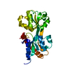
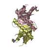
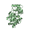
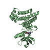
| 登録情報 | データベース: PDB / ID: 6jhk | ||||||
|---|---|---|---|---|---|---|---|
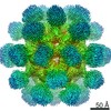
| タイトル | Crystal Structure of Bacillus subtilis RsbS | ||||||
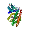
 要素 要素 | RsbS negative regulator of sigma-B | ||||||
 キーワード キーワード | PROTEIN BINDING / RsbS / stressosome / icosahedron / STAS domain | ||||||
| 機能・相同性 |  機能・相同性情報 機能・相同性情報: / STAS domain / Transcription Regulator spoIIAA / STAS domain / STAS domain profile. / STAS domain / STAS domain superfamily / 2-Layer Sandwich / Alpha Beta 類似検索 - ドメイン・相同性 | ||||||
| 生物種 |  | ||||||
| 手法 |  X線回折 / シンクロトロン / 単波長異常分散 / 解像度: 3.101 Å X線回折 / シンクロトロン / 単波長異常分散 / 解像度: 3.101 Å | ||||||
| Model details | Crystal structure of icosahedral BsRsbS | ||||||
 データ登録者 データ登録者 | Kwon, E. / Pathak, D. / Dahal, P. / Kim, D.Y. | ||||||
| 資金援助 |  韓国, 1件 韓国, 1件
| ||||||
 引用 引用 | ジャーナル: IUCrJ / 年: 2019 タイトル: Structural insights into stressosome assembly. 著者: Eunju Kwon / Deepak Pathak / Han-Ul Kim / Pawan Dahal / Sung Chul Ha / Seung Sik Lee / Hyeongseop Jeong / Dooil Jeoung / Hyeun Wook Chang / Hyun Suk Jung / Dong Young Kim / 要旨: The stressosome transduces environmental stress signals to SigB to upregulate SigB-dependent transcription, which is required for bacterial viability. The stressosome core is composed of RsbS and at ...The stressosome transduces environmental stress signals to SigB to upregulate SigB-dependent transcription, which is required for bacterial viability. The stressosome core is composed of RsbS and at least one of the RsbR paralogs. A previous cryo-electron microscopy (cryo-EM) structure of the RsbRA-RsbS complex determined under a 2 symmetry restraint showed that the stressosome core forms a pseudo-icosahedron consisting of 60 STAS domains of RsbRA and RsbS. However, it is still unclear how RsbS and one of the RsbR paralogs assemble into the stressosome. Here, an assembly model of the stressosome is presented based on the crystal structure of the RsbS icosahedron and cryo-EM structures of the RsbRA-RsbS complex determined under diverse symmetry restraints (nonsymmetric 1, dihedral 2 and icosahedral envelopes). 60 monomers of the crystal structure of RsbS fitted well into the -restrained cryo-EM structure determined at 4.1 Å resolution, even though the STAS domains in the envelope were averaged. This indicates that RsbS and RsbRA share a highly conserved STAS fold. 22 protrusions observed in the 1 envelope, corresponding to dimers of the RsbRA N-domain, allowed the STAS domains of RsbRA and RsbS to be distinguished in the stressosome core. Based on these, the model of the stressosome core was reconstructed. The mutation of RsbRA residues at the binding interface in the model (R189A/Q191A) significantly reduced the interaction between RsbRA and RsbS. These results suggest that nonconserved residues in the conserved STAS folds between RsbS and RsbR paralogs determine stressosome assembly. | ||||||
| 履歴 |
|
- 構造の表示
構造の表示
| 構造ビューア | 分子: MolmilJmol/JSmol |
|---|
- ダウンロードとリンク
-ダウンロード
| PDBx/mmCIF形式 | 6jhk.cif.gz | 119.5 KB | 表示 | PDBx/mmCIF形式 |
|---|---|---|---|---|
| PDB形式 | pdb6jhk.ent.gz | 94.8 KB | 表示 | PDB形式 |
| PDBx/mmJSON形式 | 6jhk.json.gz | ツリー表示 | PDBx/mmJSON形式 | |
| その他 |  その他のダウンロード その他のダウンロード |
-検証レポート
| 文書・要旨 | 6jhk_validation.pdf.gz | 469.3 KB | 表示 | wwPDB検証レポート |
|---|---|---|---|---|
| 文書・詳細版 | 6jhk_full_validation.pdf.gz | 478.9 KB | 表示 | |
| XML形式データ | 6jhk_validation.xml.gz | 22.8 KB | 表示 | |
| CIF形式データ | 6jhk_validation.cif.gz | 29.9 KB | 表示 | |
| アーカイブディレクトリ | https://data.pdbj.org/pub/pdb/validation_reports/jh/6jhkftp://data.pdbj.org/pub/pdb/validation_reports/jh/6jhk | HTTPS FTP |
-関連構造データ


-リンク
 PDBj
PDBj
- 集合体
集合体
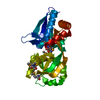
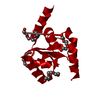
| 登録構造単位 | 
| ||||||||
|---|---|---|---|---|---|---|---|---|---|
| 1 | 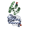
| ||||||||
| 2 | 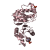
| ||||||||
| 3 | 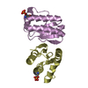
| ||||||||
| 4 |
| ||||||||
| 単位格子 |
|
-要素
| #1: タンパク質 | 分子量: 13544.638 Da / 分子数: 5 / 由来タイプ: 組換発現 / 由来: (組換発現) 遺伝子: B4122_0858, B4417_4413, CJ481_02205, ETA10_02695, ETK61_02665, ETK71_02600 プラスミド: pETDuet-1 / 発現宿主: Has protein modification | Y | |
|---|
-実験情報
-実験
| 実験 | 手法: X線回折 / 使用した結晶の数: 1 |
|---|
- 試料調製
試料調製
| 結晶 | マシュー密度: 3.75 Å3/Da / 溶媒含有率: 67.2 % |
|---|---|
| 結晶化 | 温度: 295 K / 手法: batch mode / pH: 6 / 詳細: MPD, LiSO4 |
-データ収集
| 回折 | 平均測定温度: 100 K / Serial crystal experiment: N |
|---|---|
| 放射光源 | 由来: シンクロトロン / サイト: PAL/PLS / ビームライン: 7A (6B, 6C1) / 波長: 0.97933 Å |
| 検出器 | タイプ: ADSC QUANTUM 270 / 検出器: CCD / 日付: 2016年12月15日 |
| 放射 | プロトコル: SINGLE WAVELENGTH / 単色(M)・ラウエ(L): M / 散乱光タイプ: x-ray |
| 放射波長 | 波長: 0.97933 Å / 相対比: 1 |
| 反射 | 解像度: 3.1→30 Å / Num. obs: 17310 / % possible obs: 98.9 % / 冗長度: 5.97 % / Biso Wilson estimate: 92.68 Å2 / Rmerge(I) obs: 0.072 / Net I/σ(I): 38.9 |
| 反射 シェル | 解像度: 3.1→3.15 Å / Rmerge(I) obs: 0.546 / Mean I/σ(I) obs: 5.2 / Num. unique obs: 851 / % possible all: 100 |
- 解析
解析
| ソフトウェア |
| |||||||||||||||||||||||||||||||||||||||||||||||||||||||||||||||||||||||||||||||||||||||||||
|---|---|---|---|---|---|---|---|---|---|---|---|---|---|---|---|---|---|---|---|---|---|---|---|---|---|---|---|---|---|---|---|---|---|---|---|---|---|---|---|---|---|---|---|---|---|---|---|---|---|---|---|---|---|---|---|---|---|---|---|---|---|---|---|---|---|---|---|---|---|---|---|---|---|---|---|---|---|---|---|---|---|---|---|---|---|---|---|---|---|---|---|---|
| 精密化 | 構造決定の手法: 単波長異常分散 / 解像度: 3.101→29.867 Å / SU ML: 0.34 / 交差検証法: THROUGHOUT / σ(F): 1.34 / 位相誤差: 24.37
| |||||||||||||||||||||||||||||||||||||||||||||||||||||||||||||||||||||||||||||||||||||||||||
| 溶媒の処理 | 減衰半径: 0.9 Å / VDWプローブ半径: 1.11 Å | |||||||||||||||||||||||||||||||||||||||||||||||||||||||||||||||||||||||||||||||||||||||||||
| 原子変位パラメータ | Biso max: 145.96 Å2 / Biso mean: 82.3339 Å2 / Biso min: 51.25 Å2 | |||||||||||||||||||||||||||||||||||||||||||||||||||||||||||||||||||||||||||||||||||||||||||
| 精密化ステップ | サイクル: final / 解像度: 3.101→29.867 Å
| |||||||||||||||||||||||||||||||||||||||||||||||||||||||||||||||||||||||||||||||||||||||||||
| 拘束条件 |
| |||||||||||||||||||||||||||||||||||||||||||||||||||||||||||||||||||||||||||||||||||||||||||
| LS精密化 シェル | Refine-ID: X-RAY DIFFRACTION / Rfactor Rfree error: 0 / Total num. of bins used: 12
|
