 Movie
Movie Controller


+ Open data

- Basic information
| Entry | Database: PDB / ID: 6jgy | ||||||
|---|---|---|---|---|---|---|---|
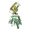
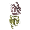
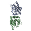
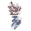
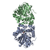
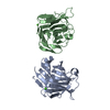
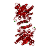
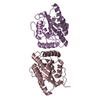
| Title | Crystal structure of LASV-GP2 in a post fusion conformation | ||||||
 Components Components | Pre-glycoprotein polyprotein GP complex | ||||||
 Keywords Keywords | VIRAL PROTEIN / LASV / GP2 | ||||||
| Function / homology |  Function and homology information Function and homology informationhost cell Golgi membrane / receptor-mediated endocytosis of virus by host cell / host cell endoplasmic reticulum membrane / fusion of virus membrane with host endosome membrane / viral envelope / virion attachment to host cell / host cell plasma membrane / virion membrane / membrane / metal ion binding Similarity search - Function | ||||||
| Biological species |  Lassa mammarenavirus Lassa mammarenavirus | ||||||
| Method |  X-RAY DIFFRACTION / SYNCHROTRON / MOLECULAR REPLACEMENT / Resolution: 3.389 Å X-RAY DIFFRACTION / SYNCHROTRON / MOLECULAR REPLACEMENT / Resolution: 3.389 Å | ||||||
 Authors Authors | Zhu, Y. / Zhang, X. / Chen, B. / Ye, S. / Zhang, R. | ||||||
 Citation Citation | Journal: Front Microbiol / Year: 2019 Title: Crystal Structure of Refolding Fusion Core of Lassa Virus GP2 and Design of Lassa Virus Fusion Inhibitors. Authors: Zhang, X. / Wang, C. / Chen, B. / Wang, Q. / Xu, W. / Ye, S. / Jiang, S. / Zhu, Y. / Zhang, R. | ||||||
| History |
|
- Structure visualization
| Structure viewer | Molecule: MolmilJmol/JSmol |
|---|
- Downloads & links
-Download
| PDBx/mmCIF format | 6jgy.cif.gz | 35.4 KB | Display | PDBx/mmCIF format |
|---|---|---|---|---|
| PDB format | pdb6jgy.ent.gz | 23 KB | Display | PDB format |
| PDBx/mmJSON format | 6jgy.json.gz | Tree view | PDBx/mmJSON format | |
| Others |  Other downloads Other downloads |
-Validation report
| Arichive directory | https://data.pdbj.org/pub/pdb/validation_reports/jg/6jgyftp://data.pdbj.org/pub/pdb/validation_reports/jg/6jgy | HTTPS FTP |
|---|
-Related structure data
| Related structure data |  5omiS S: Starting model for refinement |
|---|---|
| Similar structure data |


-Links
 PDBj
PDBj- Assembly
| Deposited unit | 
| ||||||||
|---|---|---|---|---|---|---|---|---|---|
| 1 | 
| ||||||||
| Unit cell |
|
-Components
| #1: Protein | Mass: 15109.193 Da / Num. of mol.: 1 Source method: isolated from a genetically manipulated source Source: (gene. exp.) Lassa mammarenavirus / Gene: GP, GPC / Production host:  |
|---|---|
| Has protein modification | Y |
-Experimental details
-Experiment
| Experiment | Method: X-RAY DIFFRACTION / Number of used crystals: 1 |
|---|
- Sample preparation
| Crystal | Density Matthews: 3.15 Å3/Da / Density % sol: 60.9 % |
|---|---|
| Crystal grow | Temperature: 289 K / Method: vapor diffusion, hanging drop Details: 0.17 M Ammonium Acetate, 0.085 M Sodium Citrate:HCl, pH 5.6, 25.5% (w/v) PEG 4000, 15% (v/v) Glycerol |
-Data collection
| Diffraction | Mean temperature: 100 K / Serial crystal experiment: N |
|---|---|
| Diffraction source | Source: SYNCHROTRON / Site: SSRF  / Beamline: BL18U1 / Wavelength: 0.9793 Å / Beamline: BL18U1 / Wavelength: 0.9793 Å |
| Detector | Type: DECTRIS PILATUS 2M / Detector: PIXEL / Date: May 27, 2018 |
| Radiation | Protocol: SINGLE WAVELENGTH / Monochromatic (M) / Laue (L): M / Scattering type: x-ray |
| Radiation wavelength | Wavelength: 0.9793 Å / Relative weight: 1 |
| Reflection | Resolution: 3.389→38.037 Å / Num. obs: 2839 / % possible obs: 98.1 % / Redundancy: 6.3 % / Net I/σ(I): 19.15 |
| Reflection shell | Resolution: 3.4→3.48 Å |
- Processing
| Software |
| ||||||||||||||||||||||||
|---|---|---|---|---|---|---|---|---|---|---|---|---|---|---|---|---|---|---|---|---|---|---|---|---|---|
| Refinement | Method to determine structure: MOLECULAR REPLACEMENT Starting model: 5OMI Resolution: 3.389→38.037 Å / SU ML: 0.19 / Cross valid method: FREE R-VALUE / σ(F): 1.38 / Phase error: 19.8
| ||||||||||||||||||||||||
| Solvent computation | Shrinkage radii: 0.9 Å / VDW probe radii: 1.11 Å | ||||||||||||||||||||||||
| Refinement step | Cycle: LAST / Resolution: 3.389→38.037 Å
| ||||||||||||||||||||||||
| Refine LS restraints |
| ||||||||||||||||||||||||
| LS refinement shell | Highest resolution: 3.389 Å
|
