Protocol: SINGLE WAVELENGTH / Monochromatic (M) / Laue (L): M / Scattering type: x-ray
Radiation wavelength
Wavelength: 0.979 Å / Relative weight: 1
Reflection
Resolution: 1.18→62.21 Å / Num. obs: 171991 / % possible obs: 91.49 % / Redundancy: 3.6 % / Biso Wilson estimate: 11.45 Å2 / Rmerge(I) obs: 0.054 / Net I/σ(I): 24.5
Reflection shell
Resolution: 1.18→1.22 Å / Redundancy: 3.7 % / Rmerge(I) obs: 0.393 / Mean I/σ(I) obs: 3.5 / % possible all: 89.43
-
Processing
Software
Name
Version
Classification
HKL-2000
datareduction
HKL-2000
datascaling
REFMAC
5.8.0135
refinement
PDB_EXTRACT
3.24
dataextraction
PHASER
phasing
Refinement
Method to determine structure: MOLECULAR REPLACEMENT / Resolution: 1.18→62.21 Å / Cor.coef. Fo:Fc: 0.982 / Cor.coef. Fo:Fc free: 0.977 / SU B: 0.501 / SU ML: 0.023 / Cross valid method: THROUGHOUT / σ(F): 0 / ESU R: 0.033 / ESU R Free: 0.035 Details: 1. HYDROGENS HAVE BEEN ADDED IN THE RIDING POSITIONS U VALUES : REFINED INDIVIDUALLY 2. The unusual chirality concerning XYP 504 was due to that O4A atom of XYP 504 was captured by residue ...Details: 1. HYDROGENS HAVE BEEN ADDED IN THE RIDING POSITIONS U VALUES : REFINED INDIVIDUALLY 2. The unusual chirality concerning XYP 504 was due to that O4A atom of XYP 504 was captured by residue ARG 246 and formed 3 H-bonds. The unusual chirality concerning XYP 502 was due to that the O2B, O3B and O4B atoms were fixed by protein through 7 H-bonds.
Rfactor
Num. reflection
% reflection
Selection details
Rfree
0.151
8599
5 %
RANDOM
Rwork
0.1321
-
-
-
obs
0.133
163429
91.4 %
-
Solvent computation
Ion probe radii: 0.8 Å / Shrinkage radii: 0.8 Å / VDW probe radii: 1.2 Å
In the structure databanks used in Yorodumi, some data are registered as the other names, "COVID-19 virus" and "2019-nCoV". Here are the details of the virus and the list of structure data.
Jan 31, 2019. EMDB accession codes are about to change! (news from PDBe EMDB page)
EMDB accession codes are about to change! (news from PDBe EMDB page)
The allocation of 4 digits for EMDB accession codes will soon come to an end. Whilst these codes will remain in use, new EMDB accession codes will include an additional digit and will expand incrementally as the available range of codes is exhausted. The current 4-digit format prefixed with “EMD-” (i.e. EMD-XXXX) will advance to a 5-digit format (i.e. EMD-XXXXX), and so on. It is currently estimated that the 4-digit codes will be depleted around Spring 2019, at which point the 5-digit format will come into force.
The EM Navigator/Yorodumi systems omit the EMD- prefix.
Related info.:Q: What is EMD? / ID/Accession-code notation in Yorodumi/EM Navigator
Yorodumi is a browser for structure data from EMDB, PDB, SASBDB, etc.
This page is also the successor to EM Navigator detail page, and also detail information page/front-end page for Omokage search.
The word "yorodu" (or yorozu) is an old Japanese word meaning "ten thousand". "mi" (miru) is to see.
Related info.:EMDB / PDB / SASBDB / Comparison of 3 databanks / Yorodumi Search / Aug 31, 2016. New EM Navigator & Yorodumi / Yorodumi Papers / Jmol/JSmol / Function and homology information / Changes in new EM Navigator and Yorodumi
 Movie
Movie Controller

 Yorodumi
 Open data
 Basic information
 Components
 Keywords
 Function and homology information
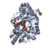
 X-RAY DIFFRACTION /
  Authors
 Citation
 Structure visualization
 Downloads & links
 Other downloads


 PDBj
PDBj
 Assembly


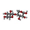

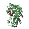
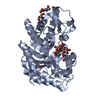
 Type: D-saccharide, beta linking / Mass: 221.208 Da / Num. of mol.: 2


 Mass: 92.094 Da / Num. of mol.: 5 / Source method: obtained synthetically / Formula: C3H8O3
 Mass: 195.237 Da / Num. of mol.: 1 / Source method: obtained synthetically / Formula: C6H13NO4S / Comment: pH buffer*YM
 Mass: 106.120 Da / Num. of mol.: 2 / Source method: obtained synthetically / Formula: C4H10O3
 Sample preparation
 / Beamline: BL19U1 / Wavelength: 0.979 Å
 Processing