 Movie
Movie Controller


+ Open data

- Basic information
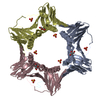
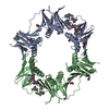
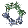
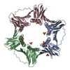
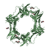
| Entry | Database: PDB / ID: 6j8y | ||||||
|---|---|---|---|---|---|---|---|
| Title | Crystal structure of the human RAD9-HUS1-RAD1-RHINO complex | ||||||
 Components Components |
| ||||||
 Keywords Keywords | CELL CYCLE / dna damage / checkpoint / dna repair / dna binding clamp / exonuclease / hydrolase / nuclease / nucleus / phosphoprotein / pcna | ||||||
| Function / homology |  Function and homology information Function and homology informationmeiotic DNA integrity checkpoint signaling / checkpoint clamp complex / meiotic recombination checkpoint signaling / positive regulation of G0 to G1 transition / exodeoxyribonuclease III / double-stranded DNA 3'-5' DNA exonuclease activity / DNA replication checkpoint signaling / 3'-5'-DNA exonuclease activity / embryo development ending in birth or egg hatching / double-strand break repair via alternative nonhomologous end joining ...meiotic DNA integrity checkpoint signaling / checkpoint clamp complex / meiotic recombination checkpoint signaling / positive regulation of G0 to G1 transition / exodeoxyribonuclease III / double-stranded DNA 3'-5' DNA exonuclease activity / DNA replication checkpoint signaling / 3'-5'-DNA exonuclease activity / embryo development ending in birth or egg hatching / double-strand break repair via alternative nonhomologous end joining / mitotic DNA replication checkpoint signaling / protein localization to site of double-strand break / chromatin-protein adaptor activity / mitotic intra-S DNA damage checkpoint signaling / positive regulation of intrinsic apoptotic signaling pathway in response to DNA damage / recombinational repair / HDR through Single Strand Annealing (SSA) / Impaired BRCA2 binding to RAD51 / Presynaptic phase of homologous DNA pairing and strand exchange / Activation of ATR in response to replication stress / response to UV / substantia nigra development / DNA damage checkpoint signaling / telomere maintenance / cellular response to ionizing radiation / nucleotide-excision repair / SH3 domain binding / G2/M DNA damage checkpoint / double-strand break repair via homologous recombination / intrinsic apoptotic signaling pathway in response to DNA damage / histone deacetylase binding / cellular response to UV / chromosome / site of double-strand break / Processing of DNA double-strand break ends / Regulation of TP53 Activity through Phosphorylation / damaged DNA binding / DNA repair / DNA damage response / protein kinase binding / chromatin / nucleolus / enzyme binding / nucleoplasm / nucleus / cytoplasm / cytosol Similarity search - Function | ||||||
| Biological species |  Homo sapiens (human) Homo sapiens (human) | ||||||
| Method |  X-RAY DIFFRACTION / SYNCHROTRON / MOLECULAR REPLACEMENT / Resolution: 2.4 Å X-RAY DIFFRACTION / SYNCHROTRON / MOLECULAR REPLACEMENT / Resolution: 2.4 Å | ||||||
 Authors Authors | Hara, K. / Iida, N. / Sakurai, H. / Hashimoto, H. | ||||||
 Citation Citation | Journal: J.Biol.Chem. / Year: 2020 Title: Structure of the RAD9-RAD1-HUS1 checkpoint clamp bound to RHINO sheds light on the other side of the DNA clamp. Authors: Hara, K. / Iida, N. / Tamafune, R. / Ohashi, E. / Sakurai, H. / Ishikawa, Y. / Hishiki, A. / Hashimoto, H. | ||||||
| History |
|
- Structure visualization
| Structure viewer | Molecule: MolmilJmol/JSmol |
|---|
- Downloads & links
-Download
| PDBx/mmCIF format | 6j8y.cif.gz | 170.9 KB | Display | PDBx/mmCIF format |
|---|---|---|---|---|
| PDB format | pdb6j8y.ent.gz | 132 KB | Display | PDB format |
| PDBx/mmJSON format | 6j8y.json.gz | Tree view | PDBx/mmJSON format | |
| Others |  Other downloads Other downloads |
-Validation report
| Arichive directory | https://data.pdbj.org/pub/pdb/validation_reports/j8/6j8yftp://data.pdbj.org/pub/pdb/validation_reports/j8/6j8y | HTTPS FTP |
|---|
-Related structure data
| Related structure data |  3a1jS S: Starting model for refinement |
|---|---|
| Similar structure data |


-Links
 PDBj
PDBj


- Assembly
| Deposited unit | 
| ||||||||
|---|---|---|---|---|---|---|---|---|---|
| 1 |
| ||||||||
| Unit cell |
|
-Components
| #1: Protein | Mass: 29746.393 Da / Num. of mol.: 1 Source method: isolated from a genetically manipulated source Source: (gene. exp.) Homo sapiens (human) / Gene: RAD9A / Production host:  |
|---|---|
| #2: Protein | Mass: 32560.734 Da / Num. of mol.: 1 Source method: isolated from a genetically manipulated source Source: (gene. exp.) Homo sapiens (human) / Gene: HUS1 / Production host: |
| #3: Protein | Mass: 31854.201 Da / Num. of mol.: 1 Source method: isolated from a genetically manipulated source Source: (gene. exp.) Homo sapiens (human) / Gene: RAD1, REC1 / Production host: |
| #4: Protein/peptide | Mass: 2206.365 Da / Num. of mol.: 1 Source method: isolated from a genetically manipulated source Source: (gene. exp.) Homo sapiens (human) / Gene: RHNO1 / Production host: |
| #5: Water | ChemComp-HOH /  Mass: 18.015 Da / Num. of mol.: 68 / Source method: isolated from a natural source / Formula: H2O Mass: 18.015 Da / Num. of mol.: 68 / Source method: isolated from a natural source / Formula: H2O |
| Has protein modification | Y |
-Experimental details
-Experiment
| Experiment | Method: X-RAY DIFFRACTION / Number of used crystals: 1 |
|---|
- Sample preparation
| Crystal | Density Matthews: 2.88 Å3/Da / Density % sol: 57.31 % |
|---|---|
| Crystal grow | Temperature: 293 K / Method: vapor diffusion, hanging drop / Details: PEG 3350, Sodium citrate, Bis-tris propane pH 7.5 |
-Data collection
| Diffraction | Mean temperature: 100 K / Serial crystal experiment: N |
|---|---|
| Diffraction source | Source: SYNCHROTRON / Site: Photon Factory  / Beamline: BL-17A / Wavelength: 0.98 Å / Beamline: BL-17A / Wavelength: 0.98 Å |
| Detector | Type: DECTRIS EIGER X 16M / Detector: PIXEL / Date: Nov 17, 2018 |
| Radiation | Protocol: SINGLE WAVELENGTH / Monochromatic (M) / Laue (L): M / Scattering type: x-ray |
| Radiation wavelength | Wavelength: 0.98 Å / Relative weight: 1 |
| Reflection | Resolution: 2.4→20 Å / Num. obs: 44421 / % possible obs: 99.6 % / Redundancy: 6.68 % / CC1/2: 0.999 / Rmerge(I) obs: 0.066176 / Net I/σ(I): 21.17 |
| Reflection shell | Resolution: 2.4→2.53 Å / Rmerge(I) obs: 0.7791 / Num. unique obs: 27653 / CC1/2: 0.742 |
- Processing
| Software |
| |||||||||||||||||||||||||||||||||||||||||||||||||||||||||||||||||||||||||||||||||||||||||||||||||||||||||||||||||||||||
|---|---|---|---|---|---|---|---|---|---|---|---|---|---|---|---|---|---|---|---|---|---|---|---|---|---|---|---|---|---|---|---|---|---|---|---|---|---|---|---|---|---|---|---|---|---|---|---|---|---|---|---|---|---|---|---|---|---|---|---|---|---|---|---|---|---|---|---|---|---|---|---|---|---|---|---|---|---|---|---|---|---|---|---|---|---|---|---|---|---|---|---|---|---|---|---|---|---|---|---|---|---|---|---|---|---|---|---|---|---|---|---|---|---|---|---|---|---|---|---|---|
| Refinement | Method to determine structure: MOLECULAR REPLACEMENT Starting model: 3A1J Resolution: 2.4→19.802 Å / SU ML: 0.32 / Cross valid method: FREE R-VALUE / σ(F): 1.34 / Phase error: 28.97
| |||||||||||||||||||||||||||||||||||||||||||||||||||||||||||||||||||||||||||||||||||||||||||||||||||||||||||||||||||||||
| Solvent computation | Shrinkage radii: 0.9 Å / VDW probe radii: 1.11 Å | |||||||||||||||||||||||||||||||||||||||||||||||||||||||||||||||||||||||||||||||||||||||||||||||||||||||||||||||||||||||
| Refinement step | Cycle: LAST / Resolution: 2.4→19.802 Å
| |||||||||||||||||||||||||||||||||||||||||||||||||||||||||||||||||||||||||||||||||||||||||||||||||||||||||||||||||||||||
| Refine LS restraints |
| |||||||||||||||||||||||||||||||||||||||||||||||||||||||||||||||||||||||||||||||||||||||||||||||||||||||||||||||||||||||
| LS refinement shell |
|
