 Movie
Movie Controller

[English] 日本語
 Yorodumi
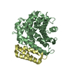
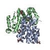
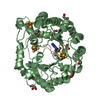
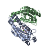
- PDB-6j80: Human mitochondrial Oligoribonuclease in complex with poly-dT DNA -
+ Open data

- Basic information
| Entry | Database: PDB / ID: 6j80 | ||||||
|---|---|---|---|---|---|---|---|
| Title | Human mitochondrial Oligoribonuclease in complex with poly-dT DNA | ||||||
 Components Components |
| ||||||
 Keywords Keywords | HYDROLASE/DNA / Exoribonuclease / Mitochonrial Oligoribonuclease / HYDROLASE / HYDROLASE-DNA complex | ||||||
| Function / homology |  Function and homology information Function and homology informationmitochondrial RNA surveillance / Hydrolases; Acting on ester bonds; Exonucleases that are active with either ribo- or deoxyribonucleic acids and produce 5'-phosphomonoesters / Mitochondrial RNA degradation / 3'-5'-DNA exonuclease activity / nucleotide metabolic process / nucleobase-containing compound metabolic process / 3'-5' exonuclease activity / mitochondrial intermembrane space / 3'-5'-RNA exonuclease activity / nucleic acid binding ...mitochondrial RNA surveillance / Hydrolases; Acting on ester bonds; Exonucleases that are active with either ribo- or deoxyribonucleic acids and produce 5'-phosphomonoesters / Mitochondrial RNA degradation / 3'-5'-DNA exonuclease activity / nucleotide metabolic process / nucleobase-containing compound metabolic process / 3'-5' exonuclease activity / mitochondrial intermembrane space / 3'-5'-RNA exonuclease activity / nucleic acid binding / mitochondrial matrix / focal adhesion / nucleolus / magnesium ion binding / mitochondrion / nucleus / cytoplasm Similarity search - Function | ||||||
| Biological species |  Homo sapiens (human) Homo sapiens (human)synthetic construct (others) | ||||||
| Method |  X-RAY DIFFRACTION / SYNCHROTRON / MOLECULAR REPLACEMENT / Resolution: 1.812 Å X-RAY DIFFRACTION / SYNCHROTRON / MOLECULAR REPLACEMENT / Resolution: 1.812 Å | ||||||
 Authors Authors | Chu, L.Y. / Agrawal, S. / Yuan, H.S. | ||||||
 Citation Citation | Journal: Rna / Year: 2019 Title: Structural insights into nanoRNA degradation by human Rexo2. Authors: Chu, L.Y. / Agrawal, S. / Chen, Y.P. / Yang, W.Z. / Yuan, H.S. | ||||||
| History |
|
- Structure visualization
| Structure viewer | Molecule: MolmilJmol/JSmol |
|---|
- Downloads & links
-Download
| PDBx/mmCIF format | 6j80.cif.gz | 89 KB | Display | PDBx/mmCIF format |
|---|---|---|---|---|
| PDB format | pdb6j80.ent.gz | 64.3 KB | Display | PDB format |
| PDBx/mmJSON format | 6j80.json.gz | Tree view | PDBx/mmJSON format | |
| Others |  Other downloads Other downloads |
-Validation report
| Arichive directory | https://data.pdbj.org/pub/pdb/validation_reports/j8/6j80ftp://data.pdbj.org/pub/pdb/validation_reports/j8/6j80 | HTTPS FTP |
|---|
-Related structure data
| Related structure data | 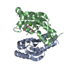 6j7yC 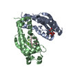 6j7zC 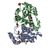 2igiS S: Starting model for refinement C: citing same article ( |
|---|---|
| Similar structure data |


-Links
 PDBj
PDBj- Assembly
| Deposited unit | 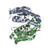
| ||||||||
|---|---|---|---|---|---|---|---|---|---|
| 1 |
| ||||||||
| Unit cell |
|
-Components
| #1: Protein | Mass: 20952.057 Da / Num. of mol.: 2 / Mutation: D199A Source method: isolated from a genetically manipulated source Source: (gene. exp.) Homo sapiens (human) / Production host:  #2: DNA chain | | Mass: 2084.392 Da / Num. of mol.: 1 / Source method: obtained synthetically / Source: (synth.) synthetic construct (others) #3: Chemical | ChemComp-CIT / |   Mass: 192.124 Da / Num. of mol.: 1 / Source method: obtained synthetically / Formula: C6H8O7 Mass: 192.124 Da / Num. of mol.: 1 / Source method: obtained synthetically / Formula: C6H8O7#4: Chemical |   Mass: 24.305 Da / Num. of mol.: 2 / Source method: obtained synthetically / Formula: Mg Mass: 24.305 Da / Num. of mol.: 2 / Source method: obtained synthetically / Formula: Mg#5: Water | ChemComp-HOH / |  Mass: 18.015 Da / Num. of mol.: 201 / Source method: isolated from a natural source / Formula: H2O Mass: 18.015 Da / Num. of mol.: 201 / Source method: isolated from a natural source / Formula: H2O |
|---|
-Experimental details
-Experiment
| Experiment | Method: X-RAY DIFFRACTION / Number of used crystals: 1 |
|---|
- Sample preparation
| Crystal | Density Matthews: 2.9 Å3/Da / Density % sol: 62.05 % |
|---|---|
| Crystal grow | Temperature: 298.15 K / Method: vapor diffusion, hanging drop / Details: 0.2 M Ammonium citrate dibasic, 20% w/v PEG 3350 |
-Data collection
| Diffraction | Mean temperature: 100 K / Serial crystal experiment: N | |||||||||||||||||||||||||||||||||||||||||||||||||||||||||||||||||||||||||||||||||||||||||||||||||||||||||||||||||||||||||||||||||||||||||||||||||||
|---|---|---|---|---|---|---|---|---|---|---|---|---|---|---|---|---|---|---|---|---|---|---|---|---|---|---|---|---|---|---|---|---|---|---|---|---|---|---|---|---|---|---|---|---|---|---|---|---|---|---|---|---|---|---|---|---|---|---|---|---|---|---|---|---|---|---|---|---|---|---|---|---|---|---|---|---|---|---|---|---|---|---|---|---|---|---|---|---|---|---|---|---|---|---|---|---|---|---|---|---|---|---|---|---|---|---|---|---|---|---|---|---|---|---|---|---|---|---|---|---|---|---|---|---|---|---|---|---|---|---|---|---|---|---|---|---|---|---|---|---|---|---|---|---|---|---|---|---|
| Diffraction source | Source: SYNCHROTRON / Site: NSRRC  / Beamline: BL15A1 / Wavelength: 1 Å / Beamline: BL15A1 / Wavelength: 1 Å | |||||||||||||||||||||||||||||||||||||||||||||||||||||||||||||||||||||||||||||||||||||||||||||||||||||||||||||||||||||||||||||||||||||||||||||||||||
| Detector | Type: RAYONIX MX300HE / Detector: CCD / Date: Jul 6, 2016 | |||||||||||||||||||||||||||||||||||||||||||||||||||||||||||||||||||||||||||||||||||||||||||||||||||||||||||||||||||||||||||||||||||||||||||||||||||
| Radiation | Protocol: SINGLE WAVELENGTH / Monochromatic (M) / Laue (L): M / Scattering type: x-ray | |||||||||||||||||||||||||||||||||||||||||||||||||||||||||||||||||||||||||||||||||||||||||||||||||||||||||||||||||||||||||||||||||||||||||||||||||||
| Radiation wavelength | Wavelength: 1 Å / Relative weight: 1 | |||||||||||||||||||||||||||||||||||||||||||||||||||||||||||||||||||||||||||||||||||||||||||||||||||||||||||||||||||||||||||||||||||||||||||||||||||
| Reflection | Resolution: 1.81→50 Å / Num. obs: 45376 / % possible obs: 99.4 % / Redundancy: 3.6 % / Biso Wilson estimate: 18.75 Å2 / Rmerge(I) obs: 0.027 / Χ2: 1.929 / Net I/σ(I): 38.3 | |||||||||||||||||||||||||||||||||||||||||||||||||||||||||||||||||||||||||||||||||||||||||||||||||||||||||||||||||||||||||||||||||||||||||||||||||||
| Reflection shell |
|
- Processing
| Software |
| |||||||||||||||||||||||||||||||||||||||||||||||||||||||||||||||||||||||||||||||||||||||||||||||||||||||||
|---|---|---|---|---|---|---|---|---|---|---|---|---|---|---|---|---|---|---|---|---|---|---|---|---|---|---|---|---|---|---|---|---|---|---|---|---|---|---|---|---|---|---|---|---|---|---|---|---|---|---|---|---|---|---|---|---|---|---|---|---|---|---|---|---|---|---|---|---|---|---|---|---|---|---|---|---|---|---|---|---|---|---|---|---|---|---|---|---|---|---|---|---|---|---|---|---|---|---|---|---|---|---|---|---|---|---|
| Refinement | Method to determine structure: MOLECULAR REPLACEMENT Starting model: 2IGI Resolution: 1.812→26.115 Å / SU ML: 0.15 / Cross valid method: THROUGHOUT / σ(F): 1.34 / Phase error: 21.06
| |||||||||||||||||||||||||||||||||||||||||||||||||||||||||||||||||||||||||||||||||||||||||||||||||||||||||
| Solvent computation | Shrinkage radii: 0.9 Å / VDW probe radii: 1.11 Å | |||||||||||||||||||||||||||||||||||||||||||||||||||||||||||||||||||||||||||||||||||||||||||||||||||||||||
| Displacement parameters | Biso max: 82.38 Å2 / Biso mean: 24.9434 Å2 / Biso min: 8.32 Å2 | |||||||||||||||||||||||||||||||||||||||||||||||||||||||||||||||||||||||||||||||||||||||||||||||||||||||||
| Refinement step | Cycle: final / Resolution: 1.812→26.115 Å
| |||||||||||||||||||||||||||||||||||||||||||||||||||||||||||||||||||||||||||||||||||||||||||||||||||||||||
| LS refinement shell | Refine-ID: X-RAY DIFFRACTION / Rfactor Rfree error: 0 / Total num. of bins used: 14
|
