 Movie
Movie Controller

[English] 日本語
 Yorodumi
- PDB-6j7u: Crystal structure of blue fluorescent protein from metagenomic li... -
+ Open data

- Basic information
| Entry | Database: PDB / ID: 6j7u | ||||||
|---|---|---|---|---|---|---|---|
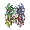
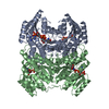
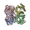
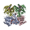
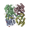
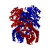
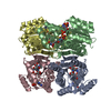
| Title | Crystal structure of blue fluorescent protein from metagenomic library in complex with NADPH | ||||||
 Components Components | Blue fluorescent protein | ||||||
 Keywords Keywords | OXIDOREDUCTASE / DEHYDROGENASE / BFP | ||||||
| Function / homology |  Function and homology information Function and homology information3-methyl-2-oxobutanoate dehydrogenase (2-methylpropanoyl-transferring) / branched-chain 2-oxo acid dehydrogenase activity / nucleotide binding Similarity search - Function | ||||||
| Biological species |  uncultured bacterium (environmental samples) uncultured bacterium (environmental samples) | ||||||
| Method |  X-RAY DIFFRACTION / SYNCHROTRON / MOLECULAR REPLACEMENT / Resolution: 2.302 Å X-RAY DIFFRACTION / SYNCHROTRON / MOLECULAR REPLACEMENT / Resolution: 2.302 Å | ||||||
 Authors Authors | Seo, P.W. / Kim, J.S. | ||||||
| Funding support |  Korea, Republic Of, 1items Korea, Republic Of, 1items
| ||||||
 Citation Citation | Journal: J.Mol.Biol. / Year: 2019 Title: Structure-Guided Generation of a Redox-Independent Blue Fluorescent Protein from mBFP. Authors: Seo, P.W. / Jo, E.S. / You, S.H. / Cheong, D.E. / Kim, G.J. / Kim, J.S. | ||||||
| History |
|
- Structure visualization
| Structure viewer | Molecule: MolmilJmol/JSmol |
|---|
- Downloads & links
-Download
| PDBx/mmCIF format | 6j7u.cif.gz | 375.7 KB | Display | PDBx/mmCIF format |
|---|---|---|---|---|
| PDB format | pdb6j7u.ent.gz | 307.8 KB | Display | PDB format |
| PDBx/mmJSON format | 6j7u.json.gz | Tree view | PDBx/mmJSON format | |
| Others |  Other downloads Other downloads |
-Validation report
| Arichive directory | https://data.pdbj.org/pub/pdb/validation_reports/j7/6j7uftp://data.pdbj.org/pub/pdb/validation_reports/j7/6j7u | HTTPS FTP |
|---|
-Related structure data
| Related structure data |  6j7hC 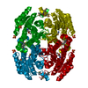 3v2gS S: Starting model for refinement C: citing same article ( |
|---|---|
| Similar structure data |


-Links
 PDBj
PDBj


- Assembly
| Deposited unit | 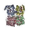
| ||||||||
|---|---|---|---|---|---|---|---|---|---|
| 1 |
| ||||||||
| Unit cell |
|
-Components
| #1: Protein | Mass: 26882.391 Da / Num. of mol.: 4 Source method: isolated from a genetically manipulated source Source: (gene. exp.) uncultured bacterium (environmental samples)Production host: References: UniProt: D6NKF4, 3-methyl-2-oxobutanoate dehydrogenase (2-methylpropanoyl-transferring) #2: Chemical | ChemComp-NDP /   Mass: 745.421 Da / Num. of mol.: 4 / Source method: obtained synthetically / Formula: C21H30N7O17P3 Mass: 745.421 Da / Num. of mol.: 4 / Source method: obtained synthetically / Formula: C21H30N7O17P3#3: Water | ChemComp-HOH / |  Mass: 18.015 Da / Num. of mol.: 231 / Source method: isolated from a natural source / Formula: H2O Mass: 18.015 Da / Num. of mol.: 231 / Source method: isolated from a natural source / Formula: H2O |
|---|
-Experimental details
-Experiment
| Experiment | Method: X-RAY DIFFRACTION / Number of used crystals: 1 |
|---|
- Sample preparation
| Crystal | Density Matthews: 2.08 Å3/Da / Density % sol: 40.81 % |
|---|---|
| Crystal grow | Temperature: 294 K / Method: vapor diffusion, hanging drop / pH: 9 Details: 0.1M BICINE pH 9.0, 0.3M Magnesium nitrate, 22% w/v PEG 2000, 4% v/v MPD |
-Data collection
| Diffraction | Mean temperature: 100 K / Serial crystal experiment: N |
|---|---|
| Diffraction source | Source: SYNCHROTRON / Site: PAL/PLS / Beamline: 11C / Wavelength: 0.9793 Å |
| Detector | Type: DECTRIS PILATUS 6M / Detector: PIXEL / Date: Dec 30, 2017 |
| Radiation | Protocol: SINGLE WAVELENGTH / Monochromatic (M) / Laue (L): M / Scattering type: x-ray |
| Radiation wavelength | Wavelength: 0.9793 Å / Relative weight: 1 |
| Reflection | Resolution: 2.3→20 Å / Num. obs: 40297 / % possible obs: 99.5 % / Redundancy: 7.4 % / Rpim(I) all: 0.07 / Net I/σ(I): 8.5 |
| Reflection shell | Resolution: 2.3→2.34 Å / Num. unique obs: 1987 / Rpim(I) all: 0.296 |
- Processing
| Software |
| ||||||||||||||||||||||||||||||||||||||||||||||||||||||||||||||||||||||||||||||||||||
|---|---|---|---|---|---|---|---|---|---|---|---|---|---|---|---|---|---|---|---|---|---|---|---|---|---|---|---|---|---|---|---|---|---|---|---|---|---|---|---|---|---|---|---|---|---|---|---|---|---|---|---|---|---|---|---|---|---|---|---|---|---|---|---|---|---|---|---|---|---|---|---|---|---|---|---|---|---|---|---|---|---|---|---|---|---|
| Refinement | Method to determine structure: MOLECULAR REPLACEMENT Starting model: 3V2G Resolution: 2.302→19.777 Å / SU ML: 0.3 / Cross valid method: FREE R-VALUE / σ(F): 1.34 / Phase error: 31.81
| ||||||||||||||||||||||||||||||||||||||||||||||||||||||||||||||||||||||||||||||||||||
| Solvent computation | Shrinkage radii: 0.9 Å / VDW probe radii: 1.11 Å | ||||||||||||||||||||||||||||||||||||||||||||||||||||||||||||||||||||||||||||||||||||
| Refinement step | Cycle: LAST / Resolution: 2.302→19.777 Å
| ||||||||||||||||||||||||||||||||||||||||||||||||||||||||||||||||||||||||||||||||||||
| Refine LS restraints |
| ||||||||||||||||||||||||||||||||||||||||||||||||||||||||||||||||||||||||||||||||||||
| LS refinement shell |
| ||||||||||||||||||||||||||||||||||||||||||||||||||||||||||||||||||||||||||||||||||||
| Refinement TLS params. | Method: refined / Origin x: 14.3715 Å / Origin y: 0.0367 Å / Origin z: -19.5246 Å
| ||||||||||||||||||||||||||||||||||||||||||||||||||||||||||||||||||||||||||||||||||||
| Refinement TLS group | Selection details: all |
