| Entry | Database: PDB / ID: 6rx5
|
|---|
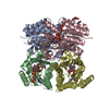
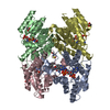
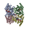
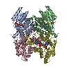
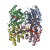
| Title | Trypanosoma brucei PTR1 (TbPTR1) in complex with inhibitor 1 (NMT-C0003) |
|---|
 Components Components | Pteridine reductase |
|---|
 Keywords Keywords | OXIDOREDUCTASE / Trypanosoma brucei / pteridine reductase / PTR1 / TbPTR1 / pteridine based inhibitors |
|---|
| Function / homology |  Function and homology information Function and homology information
Pteridine reductase / Short-chain dehydrogenase/reductase, conserved site / Short-chain dehydrogenases/reductases family signature. / Enoyl-(Acyl carrier protein) reductase / Short-chain dehydrogenase/reductase SDR / NAD(P)-binding Rossmann-like Domain / NAD(P)-binding domain superfamily / Rossmann fold / 3-Layer(aba) Sandwich / Alpha BetaSimilarity search - Domain/homology |
|---|
| Biological species |   Trypanosoma brucei brucei (eukaryote) Trypanosoma brucei brucei (eukaryote) |
|---|
| Method |  X-RAY DIFFRACTION / SYNCHROTRON / MOLECULAR REPLACEMENT / Resolution: 1.42 Å X-RAY DIFFRACTION / SYNCHROTRON / MOLECULAR REPLACEMENT / Resolution: 1.42 Å |
|---|
 Authors Authors | Landi, G. / Pozzi, C. / Mangani, S. |
|---|
| Funding support | 1items | Organization | Grant number | Country |
|---|
| European Union | 603240 | |
|
|---|
 Citation Citation | Journal: J.Med.Chem. / Year: 2022
Title: Multitarget, Selective Compound Design Yields Potent Inhibitors of a Kinetoplastid Pteridine Reductase 1.
Authors: Pohner, I. / Quotadamo, A. / Panecka-Hofman, J. / Luciani, R. / Santucci, M. / Linciano, P. / Landi, G. / Di Pisa, F. / Dello Iacono, L. / Pozzi, C. / Mangani, S. / Gul, S. / Witt, G. / ...Authors: Pohner, I. / Quotadamo, A. / Panecka-Hofman, J. / Luciani, R. / Santucci, M. / Linciano, P. / Landi, G. / Di Pisa, F. / Dello Iacono, L. / Pozzi, C. / Mangani, S. / Gul, S. / Witt, G. / Ellinger, B. / Kuzikov, M. / Santarem, N. / Cordeiro-da-Silva, A. / Costi, M.P. / Venturelli, A. / Wade, R.C. |
|---|
| History | | Deposition | Jun 7, 2019 | Deposition site: PDBE / Processing site: PDBE |
|---|
| Revision 1.0 | Jul 15, 2020 | Provider: repository / Type: Initial release |
|---|
| Revision 1.1 | Sep 13, 2023 | Group: Data collection / Database references
Category: chem_comp_atom / chem_comp_bond ...chem_comp_atom / chem_comp_bond / citation / citation_author / database_2
Item: _citation.country / _citation.journal_abbrev ..._citation.country / _citation.journal_abbrev / _citation.journal_id_ASTM / _citation.journal_id_CSD / _citation.journal_id_ISSN / _citation.journal_volume / _citation.page_first / _citation.page_last / _citation.pdbx_database_id_DOI / _citation.pdbx_database_id_PubMed / _citation.title / _citation.year / _database_2.pdbx_DOI / _database_2.pdbx_database_accession |
|---|
| Revision 1.2 | Jan 24, 2024 | Group: Refinement description / Category: pdbx_initial_refinement_model |
|---|
|
|---|
 Movie
Movie Controller

 Yorodumi
 Open data
 Basic information
 Structure visualization
 Downloads & links
 Other downloads
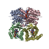


 PDBj
PDBj


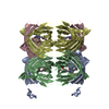
 Assembly


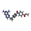


 Mass: 743.405 Da / Num. of mol.: 4 / Source method: obtained synthetically / Formula: C21H28N7O17P3
 Mass: 450.494 Da / Num. of mol.: 3 / Source method: obtained synthetically / Formula: C22H26N8O3 / Feature type: SUBJECT OF INVESTIGATION
 Mass: 92.094 Da / Num. of mol.: 1 / Source method: obtained synthetically / Formula: C3H8O3
 Mass: 59.044 Da / Num. of mol.: 1 / Source method: obtained synthetically / Formula: C2H3O2
 Sample preparation
 / Beamline: I04-1 / Wavelength: 0.91731 Å
 Processing