 Movie
Movie Controller


+ Open data

- Basic information
| Entry | Database: PDB / ID: 6j6x | ||||||||||||
|---|---|---|---|---|---|---|---|---|---|---|---|---|---|
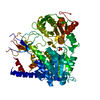
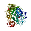
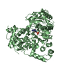
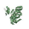
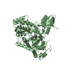
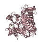
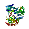
| Title | Crystal structure of apo GGTaseIII | ||||||||||||
 Components Components |
| ||||||||||||
 Keywords Keywords | LIPID BINDING PROTEIN / lipid transferase | ||||||||||||
| Function / homology |  Function and homology information Function and homology informationprotein prenyltransferase activity / protein geranylgeranyltransferase type II / Rab-protein geranylgeranyltransferase complex / Rab geranylgeranyltransferase activity / protein geranylgeranylation / RAB geranylgeranylation / TP53 regulates transcription of several additional cell death genes whose specific roles in p53-dependent apoptosis remain uncertain / endoplasmic reticulum to Golgi vesicle-mediated transport / visual perception / protein modification process ...protein prenyltransferase activity / protein geranylgeranyltransferase type II / Rab-protein geranylgeranyltransferase complex / Rab geranylgeranyltransferase activity / protein geranylgeranylation / RAB geranylgeranylation / TP53 regulates transcription of several additional cell death genes whose specific roles in p53-dependent apoptosis remain uncertain / endoplasmic reticulum to Golgi vesicle-mediated transport / visual perception / protein modification process / small GTPase binding / mitochondrion / zinc ion binding / plasma membrane / cytoplasm / cytosol Similarity search - Function | ||||||||||||
| Biological species |  Homo sapiens (human) Homo sapiens (human) | ||||||||||||
| Method |  X-RAY DIFFRACTION / SYNCHROTRON / MOLECULAR REPLACEMENT / Resolution: 2.962 Å X-RAY DIFFRACTION / SYNCHROTRON / MOLECULAR REPLACEMENT / Resolution: 2.962 Å | ||||||||||||
 Authors Authors | Goto-Ito, S. / Yamagata, A. / Sato, Y. / Fukai, S. | ||||||||||||
| Funding support |  Japan, 3items Japan, 3items
| ||||||||||||
 Citation Citation | Journal: Embo J. / Year: 2020 Title: A SNARE geranylgeranyltransferase essential for the organization of the Golgi apparatus. Authors: Shirakawa, R. / Goto-Ito, S. / Goto, K. / Wakayama, S. / Kubo, H. / Sakata, N. / Trinh, D.A. / Yamagata, A. / Sato, Y. / Masumoto, H. / Cheng, J. / Fujimoto, T. / Fukai, S. / Horiuchi, H. | ||||||||||||
| History |
|
- Structure visualization
| Structure viewer | Molecule: MolmilJmol/JSmol |
|---|
- Downloads & links
-Download
| PDBx/mmCIF format | 6j6x.cif.gz | 132.6 KB | Display | PDBx/mmCIF format |
|---|---|---|---|---|
| PDB format | pdb6j6x.ent.gz | 99.9 KB | Display | PDB format |
| PDBx/mmJSON format | 6j6x.json.gz | Tree view | PDBx/mmJSON format | |
| Others |  Other downloads Other downloads |
-Validation report
| Arichive directory | https://data.pdbj.org/pub/pdb/validation_reports/j6/6j6xftp://data.pdbj.org/pub/pdb/validation_reports/j6/6j6x | HTTPS FTP |
|---|
-Related structure data
| Related structure data |  6j74C  6j7fC  6j7xC  1dceS  3dssS S: Starting model for refinement C: citing same article ( |
|---|---|
| Similar structure data |


-Links
 PDBj
PDBj


- Assembly
| Deposited unit | 
| ||||||||
|---|---|---|---|---|---|---|---|---|---|
| 1 |
| ||||||||
| Unit cell |
|
-Components
| #1: Protein | Mass: 42228.938 Da / Num. of mol.: 1 Source method: isolated from a genetically manipulated source Source: (gene. exp.) Homo sapiens (human) / Gene: PTAR1 / Production host:  | ||||
|---|---|---|---|---|---|
| #2: Protein | Mass: 37371.723 Da / Num. of mol.: 1 Source method: isolated from a genetically manipulated source Source: (gene. exp.) Homo sapiens (human) / Gene: RABGGTB, GGTB / Production host: References: UniProt: P53611, protein geranylgeranyltransferase type II | ||||
| #3: Chemical |   Mass: 118.174 Da / Num. of mol.: 2 / Source method: obtained synthetically / Formula: C6H14O2 / Comment: precipitant*YM Mass: 118.174 Da / Num. of mol.: 2 / Source method: obtained synthetically / Formula: C6H14O2 / Comment: precipitant*YM#4: Chemical | ChemComp-MG / |   Mass: 24.305 Da / Num. of mol.: 1 / Source method: obtained synthetically / Formula: Mg Mass: 24.305 Da / Num. of mol.: 1 / Source method: obtained synthetically / Formula: MgHas protein modification | Y | |
-Experimental details
-Experiment
| Experiment | Method: X-RAY DIFFRACTION / Number of used crystals: 1 |
|---|
- Sample preparation
| Crystal | Density Matthews: 4.6 Å3/Da / Density % sol: 77.69 % |
|---|---|
| Crystal grow | Temperature: 293 K / Method: vapor diffusion, sitting drop Details: 0.04M MgCl2, 0.05M NaCacodylate pH 6, 5% MPD, 8mM CHAPS |
-Data collection
| Diffraction | Mean temperature: 100 K / Serial crystal experiment: N |
|---|---|
| Diffraction source | Source: SYNCHROTRON / Site: SPring-8 / Beamline: BL41XU / Wavelength: 1.044 Å |
| Detector | Type: DECTRIS PILATUS3 S 6M / Detector: PIXEL / Date: Jun 15, 2017 |
| Radiation | Protocol: SINGLE WAVELENGTH / Monochromatic (M) / Laue (L): M / Scattering type: x-ray |
| Radiation wavelength | Wavelength: 1.044 Å / Relative weight: 1 |
| Reflection | Resolution: 2.96→50 Å / Num. obs: 33185 / % possible obs: 100 % / Redundancy: 69.8 % / Rmerge(I) obs: 0.204 / Net I/σ(I): 29 |
| Reflection shell | Resolution: 2.96→3.01 Å / Redundancy: 59.5 % / Rmerge(I) obs: 1.848 / Num. unique obs: 1568 / CC1/2: 0.578 / % possible all: 100 |
- Processing
| Software |
| ||||||||||||||||||||||||||||||||||||||||||||||||||||||||||||||||||||||||||||||||||||
|---|---|---|---|---|---|---|---|---|---|---|---|---|---|---|---|---|---|---|---|---|---|---|---|---|---|---|---|---|---|---|---|---|---|---|---|---|---|---|---|---|---|---|---|---|---|---|---|---|---|---|---|---|---|---|---|---|---|---|---|---|---|---|---|---|---|---|---|---|---|---|---|---|---|---|---|---|---|---|---|---|---|---|---|---|---|
| Refinement | Method to determine structure: MOLECULAR REPLACEMENT Starting model: 3DSS, 1DCE Resolution: 2.962→49.475 Å / SU ML: 0.43 / Cross valid method: FREE R-VALUE / σ(F): 1.41 / Phase error: 29.37
| ||||||||||||||||||||||||||||||||||||||||||||||||||||||||||||||||||||||||||||||||||||
| Solvent computation | Shrinkage radii: 0.9 Å / VDW probe radii: 1.11 Å | ||||||||||||||||||||||||||||||||||||||||||||||||||||||||||||||||||||||||||||||||||||
| Refinement step | Cycle: LAST / Resolution: 2.962→49.475 Å
| ||||||||||||||||||||||||||||||||||||||||||||||||||||||||||||||||||||||||||||||||||||
| Refine LS restraints |
| ||||||||||||||||||||||||||||||||||||||||||||||||||||||||||||||||||||||||||||||||||||
| LS refinement shell |
|
