 Movie
Movie Controller


+ Open data

- Basic information
| Entry | Database: PDB / ID: 6j4e | ||||||
|---|---|---|---|---|---|---|---|
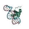
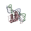
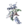
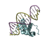
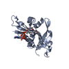
| Title | Crystal structure of the AtWRKY1 domain | ||||||
 Components Components |
| ||||||
 Keywords Keywords | DNA BINDING PROTEIN/DNA / WRKY transcription factors N-terminal WRKY domain / DNA BINDING PROTEIN / DNA BINDING PROTEIN-DNA complex | ||||||
| Function / homology |  Function and homology information Function and homology informationsalicylic acid mediated signaling pathway / response to salicylic acid / transcription cis-regulatory region binding / DNA-binding transcription factor activity / regulation of DNA-templated transcription / positive regulation of DNA-templated transcription / DNA-templated transcription / mitochondrion / DNA binding / zinc ion binding / nucleus Similarity search - Function | ||||||
| Biological species |  | ||||||
| Method |  X-RAY DIFFRACTION / SYNCHROTRON / MOLECULAR REPLACEMENT / Resolution: 3.126 Å X-RAY DIFFRACTION / SYNCHROTRON / MOLECULAR REPLACEMENT / Resolution: 3.126 Å | ||||||
 Authors Authors | Xu, Y.P. / Xu, H. / Wang, B. / Su, X.D. | ||||||
| Funding support |  China, 1items China, 1items
| ||||||
 Citation Citation | Journal: To Be Published Title: Crystal structure of the AtWRKY1 domain Authors: Xu, Y.P. / Xu, H. / Wang, B. / Su, X.D. | ||||||
| History |
|
- Structure visualization
| Structure viewer | Molecule: MolmilJmol/JSmol |
|---|
- Downloads & links
-Download
| PDBx/mmCIF format | 6j4e.cif.gz | 75.9 KB | Display | PDBx/mmCIF format |
|---|---|---|---|---|
| PDB format | pdb6j4e.ent.gz | 52.4 KB | Display | PDB format |
| PDBx/mmJSON format | 6j4e.json.gz | Tree view | PDBx/mmJSON format | |
| Others |  Other downloads Other downloads |
-Validation report
| Arichive directory | https://data.pdbj.org/pub/pdb/validation_reports/j4/6j4eftp://data.pdbj.org/pub/pdb/validation_reports/j4/6j4e | HTTPS FTP |
|---|
-Related structure data
| Related structure data |  2aydS S: Starting model for refinement |
|---|---|
| Similar structure data |


-Links
 PDBj
PDBj


- Assembly
| Deposited unit | 
| ||||||||
|---|---|---|---|---|---|---|---|---|---|
| 1 |
| ||||||||
| Unit cell |
|
-Components
| #1: DNA chain | Mass: 4569.973 Da / Num. of mol.: 1 / Source method: obtained synthetically / Source: (synth.) |
|---|---|
| #2: DNA chain | Mass: 4609.997 Da / Num. of mol.: 1 / Source method: obtained synthetically / Source: (synth.) |
| #3: Protein | Mass: 9553.848 Da / Num. of mol.: 1 Source method: isolated from a genetically manipulated source Source: (gene. exp.)  |
| #4: Chemical | ChemComp-ZN /   Mass: 65.409 Da / Num. of mol.: 1 / Source method: obtained synthetically / Formula: Zn Mass: 65.409 Da / Num. of mol.: 1 / Source method: obtained synthetically / Formula: Zn |
-Experimental details
-Experiment
| Experiment | Method: X-RAY DIFFRACTION / Number of used crystals: 1 |
|---|
- Sample preparation
| Crystal | Density Matthews: 4.33 Å3/Da / Density % sol: 71.57 % |
|---|---|
| Crystal grow | Temperature: 291 K / Method: vapor diffusion, sitting drop Details: 0.2M Ammonium sulfate, 0.1M Sodium acetate trihydrate pH 4.6, 30% Polyethylene glycol monomethyl ether 2000 |
-Data collection
| Diffraction | Mean temperature: 100 K / Serial crystal experiment: N |
|---|---|
| Diffraction source | Source: SYNCHROTRON / Site: SSRF / Beamline: BL19U1 / Wavelength: 0.978 Å |
| Detector | Type: DECTRIS PILATUS 6M / Detector: PIXEL / Date: Jul 6, 2017 |
| Radiation | Protocol: SINGLE WAVELENGTH / Monochromatic (M) / Laue (L): M / Scattering type: x-ray |
| Radiation wavelength | Wavelength: 0.978 Å / Relative weight: 1 |
| Reflection | Resolution: 3.126→28.58 Å / Num. obs: 6263 / % possible obs: 97.64 % / Redundancy: 36.8 % / CC1/2: 1 / Rmerge(I) obs: 0.1652 / Rrim(I) all: 0.1675 / Net I/σ(I): 16.02 |
| Reflection shell | Resolution: 3.126→3.238 Å / Redundancy: 37.5 % / Rmerge(I) obs: 0.5812 / Num. unique obs: 599 / CC1/2: 0.996 / Rrim(I) all: 0.5892 / % possible all: 94.02 |
- Processing
| Software |
| ||||||||||||||||||||||||||||||||||||||||
|---|---|---|---|---|---|---|---|---|---|---|---|---|---|---|---|---|---|---|---|---|---|---|---|---|---|---|---|---|---|---|---|---|---|---|---|---|---|---|---|---|---|
| Refinement | Method to determine structure: MOLECULAR REPLACEMENT Starting model: 2ayd Resolution: 3.126→28.579 Å / SU ML: 0.4 / Cross valid method: FREE R-VALUE / σ(F): 1.38 / Phase error: 23.17
| ||||||||||||||||||||||||||||||||||||||||
| Solvent computation | Shrinkage radii: 0.9 Å / VDW probe radii: 1.11 Å | ||||||||||||||||||||||||||||||||||||||||
| Refinement step | Cycle: LAST / Resolution: 3.126→28.579 Å
| ||||||||||||||||||||||||||||||||||||||||
| Refine LS restraints |
| ||||||||||||||||||||||||||||||||||||||||
| LS refinement shell |
| ||||||||||||||||||||||||||||||||||||||||
| Refinement TLS params. | Method: refined / Origin x: -46.8537 Å / Origin y: 14.9792 Å / Origin z: -15.7703 Å
| ||||||||||||||||||||||||||||||||||||||||
| Refinement TLS group | Selection details: all |
