 Movie
Movie Controller


+ Open data

- Basic information
| Entry | Database: PDB / ID: 6ime | ||||||
|---|---|---|---|---|---|---|---|
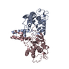
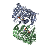
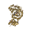
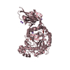
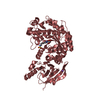
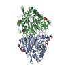
| Title | Rv2361c complex with substrate analogues | ||||||
 Components Components | Decaprenyl diphosphate synthase | ||||||
 Keywords Keywords | TRANSFERASE / decaprenyl diphosphate synthesis / substrate binding mode / catalytic mechanism / rossmann-like fold / butterfly | ||||||
| Function / homology |  Function and homology information Function and homology informationtrans,polycis-decaprenyl diphosphate synthase / Z-farnesyl diphosphate synthase activity / ditrans,polycis-polyprenyl diphosphate synthase [(2E,6E)-farnesyl diphosphate specific] / all-trans-nonaprenyl-diphosphate synthase (geranyl-diphosphate specific) activity / ditrans,polycis-undecaprenyl-diphosphate synthase [(2E,6E)-farnesyl-diphosphate specific] activity / polyprenol biosynthetic process / manganese ion binding / magnesium ion binding / plasma membrane / cytosol Similarity search - Function | ||||||
| Biological species |   Mycobacterium tuberculosis (bacteria) Mycobacterium tuberculosis (bacteria) | ||||||
| Method |  X-RAY DIFFRACTION / SYNCHROTRON / MOLECULAR REPLACEMENT / Resolution: 1.55 Å X-RAY DIFFRACTION / SYNCHROTRON / MOLECULAR REPLACEMENT / Resolution: 1.55 Å | ||||||
 Authors Authors | Ko, T.-P. / Guo, R.-T. / Chen, C.-C. / Liu, W. | ||||||
| Funding support |  Taiwan, 1items Taiwan, 1items
| ||||||
 Citation Citation | Journal: Acta Crystallogr.,Sect.F / Year: 2019 Title: Substrate-analogue complex structure of Mycobacterium tuberculosis decaprenyl diphosphate synthase. Authors: Ko, T.-P. / Xiao, X. / Guo, R.-T. / Huang, J.-W. / Liu, W. / Chen, C.-C. | ||||||
| History |
|
- Structure visualization
| Structure viewer | Molecule: MolmilJmol/JSmol |
|---|
- Downloads & links
-Download
| PDBx/mmCIF format | 6ime.cif.gz | 260.2 KB | Display | PDBx/mmCIF format |
|---|---|---|---|---|
| PDB format | pdb6ime.ent.gz | 209.4 KB | Display | PDB format |
| PDBx/mmJSON format | 6ime.json.gz | Tree view | PDBx/mmJSON format | |
| Others |  Other downloads Other downloads |
-Validation report
| Arichive directory | https://data.pdbj.org/pub/pdb/validation_reports/im/6imeftp://data.pdbj.org/pub/pdb/validation_reports/im/6ime | HTTPS FTP |
|---|
-Related structure data
| Related structure data |  4oncS S: Starting model for refinement |
|---|---|
| Similar structure data |


-Links
 PDBj
PDBj


- Assembly
| Deposited unit | 
| ||||||||
|---|---|---|---|---|---|---|---|---|---|
| 1 |
| ||||||||
| Unit cell |
|
-Components
-Protein , 1 types, 2 molecules AB
| #1: Protein | Mass: 33839.363 Da / Num. of mol.: 2 Source method: isolated from a genetically manipulated source Source: (gene. exp.) Mycobacterium tuberculosis (strain ATCC 25618 / H37Rv) (bacteria)Strain: ATCC 25618 / H37Rv / Gene: uppS, Rv2361c, MTCY27.19 / Production host: References: UniProt: P9WFF7, trans,polycis-decaprenyl diphosphate synthase, ditrans,polycis-polyprenyl diphosphate synthase [(2E,6E)-farnesyl diphosphate specific] |
|---|
-Non-polymers , 8 types, 736 molecules 


| #2: Chemical |  Mass: 330.275 Da / Num. of mol.: 2 Mass: 330.275 Da / Num. of mol.: 2Source method: isolated from a genetically manipulated source Formula: C10H20O6P2S / Feature type: SUBJECT OF INVESTIGATION #3: Chemical |  Mass: 24.305 Da / Num. of mol.: 2 / Source method: obtained synthetically / Formula: Mg Mass: 24.305 Da / Num. of mol.: 2 / Source method: obtained synthetically / Formula: Mg#4: Chemical | ChemComp-CL / |  Mass: 35.453 Da / Num. of mol.: 1 Mass: 35.453 Da / Num. of mol.: 1Source method: isolated from a genetically manipulated source Formula: Cl #5: Chemical | ChemComp-CO3 / |  Mass: 60.009 Da / Num. of mol.: 1 Mass: 60.009 Da / Num. of mol.: 1Source method: isolated from a genetically manipulated source Formula: CO3 #6: Chemical | ChemComp-DTT / |  Mass: 154.251 Da / Num. of mol.: 1 / Source method: isolated from a natural source / Formula: C4H10O2S2 Mass: 154.251 Da / Num. of mol.: 1 / Source method: isolated from a natural source / Formula: C4H10O2S2#7: Chemical | ChemComp-GOL / |  Mass: 92.094 Da / Num. of mol.: 1 / Source method: obtained synthetically / Formula: C3H8O3 Mass: 92.094 Da / Num. of mol.: 1 / Source method: obtained synthetically / Formula: C3H8O3#8: Chemical | ChemComp-ISY / |  Mass: 262.158 Da / Num. of mol.: 1 / Source method: obtained synthetically / Formula: C5H12O6P2S / Feature type: SUBJECT OF INVESTIGATION Mass: 262.158 Da / Num. of mol.: 1 / Source method: obtained synthetically / Formula: C5H12O6P2S / Feature type: SUBJECT OF INVESTIGATION#9: Water | ChemComp-HOH / | Mass: 18.015 Da / Num. of mol.: 727 / Source method: isolated from a natural source / Formula: H2O |
|---|
-Experimental details
-Experiment
| Experiment | Method: X-RAY DIFFRACTION / Number of used crystals: 1 |
|---|
- Sample preparation
| Crystal | Density Matthews: 2.52 Å3/Da / Density % sol: 51.09 % |
|---|---|
| Crystal grow | Temperature: 298 K / Method: vapor diffusion, sitting drop / pH: 7.5 Details: 0.1M HEPES, 10% GLYCEROL, 25% PEG 400, 7% PEG 3000, 1mM ISPP, 1mM GSPP |
-Data collection
| Diffraction | Mean temperature: 100 K / Serial crystal experiment: N |
|---|---|
| Diffraction source | Source: SYNCHROTRON / Site: NSRRC / Beamline: BL15A1 / Wavelength: 1 Å |
| Detector | Type: RAYONIX MX300HE / Detector: CCD / Date: Jul 3, 2015 |
| Radiation | Protocol: SINGLE WAVELENGTH / Monochromatic (M) / Laue (L): M / Scattering type: x-ray |
| Radiation wavelength | Wavelength: 1 Å / Relative weight: 1 |
| Reflection | Resolution: 1.55→25 Å / Num. obs: 95419 / % possible obs: 99.9 % / Redundancy: 6.3 % / CC1/2: 0.996 / Rmerge(I) obs: 0.036 / Rpim(I) all: 0.016 / Net I/σ(I): 48.7 |
| Reflection shell | Resolution: 1.55→1.61 Å / Redundancy: 6.3 % / Rmerge(I) obs: 0.429 / Mean I/σ(I) obs: 4.8 / Num. unique obs: 9410 / CC1/2: 0.981 / Rpim(I) all: 0.186 / % possible all: 100 |
- Processing
| Software |
| |||||||||||||||||||||||||||||||||||||||||||||||||||||||||||||||||||||||||||||||||||||||||||||||||||||||||||||||||||||||||||||||||||||||||||||||||||||||||||||||||||||||||||||||
|---|---|---|---|---|---|---|---|---|---|---|---|---|---|---|---|---|---|---|---|---|---|---|---|---|---|---|---|---|---|---|---|---|---|---|---|---|---|---|---|---|---|---|---|---|---|---|---|---|---|---|---|---|---|---|---|---|---|---|---|---|---|---|---|---|---|---|---|---|---|---|---|---|---|---|---|---|---|---|---|---|---|---|---|---|---|---|---|---|---|---|---|---|---|---|---|---|---|---|---|---|---|---|---|---|---|---|---|---|---|---|---|---|---|---|---|---|---|---|---|---|---|---|---|---|---|---|---|---|---|---|---|---|---|---|---|---|---|---|---|---|---|---|---|---|---|---|---|---|---|---|---|---|---|---|---|---|---|---|---|---|---|---|---|---|---|---|---|---|---|---|---|---|---|---|---|---|
| Refinement | Method to determine structure: MOLECULAR REPLACEMENT Starting model: 4ONC Resolution: 1.55→24.411 Å / SU ML: 0.11 / Cross valid method: THROUGHOUT / σ(F): 1.34 / Phase error: 15.63 / Stereochemistry target values: ML
| |||||||||||||||||||||||||||||||||||||||||||||||||||||||||||||||||||||||||||||||||||||||||||||||||||||||||||||||||||||||||||||||||||||||||||||||||||||||||||||||||||||||||||||||
| Solvent computation | Shrinkage radii: 0.9 Å / VDW probe radii: 1.11 Å / Solvent model: FLAT BULK SOLVENT MODEL | |||||||||||||||||||||||||||||||||||||||||||||||||||||||||||||||||||||||||||||||||||||||||||||||||||||||||||||||||||||||||||||||||||||||||||||||||||||||||||||||||||||||||||||||
| Refinement step | Cycle: LAST / Resolution: 1.55→24.411 Å
| |||||||||||||||||||||||||||||||||||||||||||||||||||||||||||||||||||||||||||||||||||||||||||||||||||||||||||||||||||||||||||||||||||||||||||||||||||||||||||||||||||||||||||||||
| Refine LS restraints |
| |||||||||||||||||||||||||||||||||||||||||||||||||||||||||||||||||||||||||||||||||||||||||||||||||||||||||||||||||||||||||||||||||||||||||||||||||||||||||||||||||||||||||||||||
| LS refinement shell |
| |||||||||||||||||||||||||||||||||||||||||||||||||||||||||||||||||||||||||||||||||||||||||||||||||||||||||||||||||||||||||||||||||||||||||||||||||||||||||||||||||||||||||||||||
| Refinement TLS params. | Method: refined / Refine-ID: X-RAY DIFFRACTION
| |||||||||||||||||||||||||||||||||||||||||||||||||||||||||||||||||||||||||||||||||||||||||||||||||||||||||||||||||||||||||||||||||||||||||||||||||||||||||||||||||||||||||||||||
| Refinement TLS group |
|
