 Movie
Movie Controller

[English] 日本語
 Yorodumi
- PDB-6ijk: Enoyl-CoA hydratase/isomerase family protein from Cupriavidus nec... -
+ Open data

- Basic information
| Entry | Database: PDB / ID: 6ijk | ||||||||||||
|---|---|---|---|---|---|---|---|---|---|---|---|---|---|
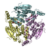
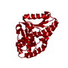
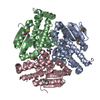
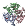
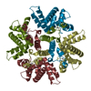
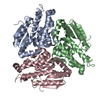
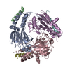
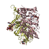
| Title | Enoyl-CoA hydratase/isomerase family protein from Cupriavidus necator H16 | ||||||||||||
 Components Components | Enoyl-CoA hydratase | ||||||||||||
 Keywords Keywords | LYASE / isomerase | ||||||||||||
| Function / homology | : / enoyl-CoA hydratase / enoyl-CoA hydratase activity / Enoyl-CoA hydratase, C-terminal / Enoyl-CoA hydratase/isomerase / Enoyl-CoA hydratase/isomerase / ClpP/crotonase-like domain superfamily / Enoyl-CoA hydratase Function and homology information Function and homology information | ||||||||||||
| Biological species |  Cupriavidus necator (bacteria) Cupriavidus necator (bacteria) | ||||||||||||
| Method |  X-RAY DIFFRACTION / SYNCHROTRON / MOLECULAR REPLACEMENT / Resolution: 2 Å X-RAY DIFFRACTION / SYNCHROTRON / MOLECULAR REPLACEMENT / Resolution: 2 Å | ||||||||||||
 Authors Authors | Seo, H. / Kim, K.-J. | ||||||||||||
| Funding support |  Korea, Republic Of, 3items Korea, Republic Of, 3items
| ||||||||||||
 Citation Citation | Journal: Biotechnol. Bioprocess Eng. / Year: 2019 Title: Crystal structure of a novel type isomerase of enoyl-CoA hydratase/isomerase family protein from Cupriavidus necator H16 Authors: Seo, H. / Kim, K.-J. | ||||||||||||
| History |
|
- Structure visualization
| Structure viewer | Molecule: MolmilJmol/JSmol |
|---|
- Downloads & links
-Download
| PDBx/mmCIF format | 6ijk.cif.gz | 113.3 KB | Display | PDBx/mmCIF format |
|---|---|---|---|---|
| PDB format | pdb6ijk.ent.gz | 86.7 KB | Display | PDB format |
| PDBx/mmJSON format | 6ijk.json.gz | Tree view | PDBx/mmJSON format | |
| Others |  Other downloads Other downloads |
-Validation report
| Arichive directory | https://data.pdbj.org/pub/pdb/validation_reports/ij/6ijkftp://data.pdbj.org/pub/pdb/validation_reports/ij/6ijk | HTTPS FTP |
|---|
-Related structure data
| Related structure data | 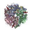 5zaiS S: Starting model for refinement |
|---|---|
| Similar structure data |
-Links
 PDBj
PDBj- Assembly
| Deposited unit | 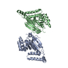
| ||||||||
|---|---|---|---|---|---|---|---|---|---|
| 1 | 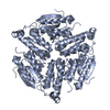
| ||||||||
| 2 | 
| ||||||||
| Unit cell |
|
-Components
| #1: Protein | Mass: 32359.475 Da / Num. of mol.: 2 Source method: isolated from a genetically manipulated source Source: (gene. exp.) Cupriavidus necator (strain ATCC 17699 / H16 / DSM 428 / Stanier 337) (bacteria)Strain: ATCC 17699 / H16 / DSM 428 / Stanier 337 / Gene: H16_B0756 / Plasmid: pET30a / Production host: #2: Water | ChemComp-HOH / |  Mass: 18.015 Da / Num. of mol.: 47 / Source method: isolated from a natural source / Formula: H2O Mass: 18.015 Da / Num. of mol.: 47 / Source method: isolated from a natural source / Formula: H2O |
|---|
-Experimental details
-Experiment
| Experiment | Method: X-RAY DIFFRACTION / Number of used crystals: 1 |
|---|
- Sample preparation
| Crystal | Density Matthews: 3.48 Å3/Da / Density % sol: 64.67 % |
|---|---|
| Crystal grow | Temperature: 293 K / Method: vapor diffusion, hanging drop / pH: 9 / Details: PEG 550 MME, NaCl, Bicine |
-Data collection
| Diffraction | Mean temperature: 100 K / Serial crystal experiment: N |
|---|---|
| Diffraction source | Source: SYNCHROTRON / Site: PAL/PLS / Beamline: 7A (6B, 6C1) / Wavelength: 0.97934 Å |
| Detector | Type: ADSC QUANTUM 270 / Detector: CCD / Date: Apr 22, 2015 |
| Radiation | Monochromator: Double Crystal Monochromator / Protocol: SINGLE WAVELENGTH / Monochromatic (M) / Laue (L): M / Scattering type: x-ray |
| Radiation wavelength | Wavelength: 0.97934 Å / Relative weight: 1 |
| Reflection | Resolution: 2→50 Å / Num. obs: 57392 / % possible obs: 99.3 % / Redundancy: 4.1 % / Rsym value: 0.09 / Net I/σ(I): 41.4 |
| Reflection shell | Resolution: 2→2.03 Å / Rmerge(I) obs: 0.32 / Mean I/σ(I) obs: 8.5 / CC1/2: 0.874 |
- Processing
| Software |
| ||||||||||||||||||||||||||||||||||||||||||||||||||||||||||||
|---|---|---|---|---|---|---|---|---|---|---|---|---|---|---|---|---|---|---|---|---|---|---|---|---|---|---|---|---|---|---|---|---|---|---|---|---|---|---|---|---|---|---|---|---|---|---|---|---|---|---|---|---|---|---|---|---|---|---|---|---|---|
| Refinement | Method to determine structure: MOLECULAR REPLACEMENT Starting model: 5ZAI Resolution: 2→35.06 Å / Cor.coef. Fo:Fc: 0.944 / Cor.coef. Fo:Fc free: 0.93 / SU B: 3.663 / SU ML: 0.101 / Cross valid method: THROUGHOUT / σ(F): 0 / ESU R: 0.136 / ESU R Free: 0.13 Details: HYDROGENS HAVE BEEN ADDED IN THE RIDING POSITIONS U VALUES : REFINED INDIVIDUALLY
| ||||||||||||||||||||||||||||||||||||||||||||||||||||||||||||
| Solvent computation | Ion probe radii: 0.8 Å / Shrinkage radii: 0.8 Å / VDW probe radii: 1.2 Å | ||||||||||||||||||||||||||||||||||||||||||||||||||||||||||||
| Displacement parameters | Biso max: 98.29 Å2 / Biso mean: 33.317 Å2 / Biso min: 19.42 Å2
| ||||||||||||||||||||||||||||||||||||||||||||||||||||||||||||
| Refinement step | Cycle: final / Resolution: 2→35.06 Å
| ||||||||||||||||||||||||||||||||||||||||||||||||||||||||||||
| Refine LS restraints |
| ||||||||||||||||||||||||||||||||||||||||||||||||||||||||||||
| LS refinement shell | Resolution: 2→2.052 Å / Rfactor Rfree error: 0 / Total num. of bins used: 20
|
