 Movie
Movie Controller


+ Open data

- Basic information
| Entry | Database: PDB / ID: 6igz | ||||||||||||||||||||||||||||||||||||||||||||||||
|---|---|---|---|---|---|---|---|---|---|---|---|---|---|---|---|---|---|---|---|---|---|---|---|---|---|---|---|---|---|---|---|---|---|---|---|---|---|---|---|---|---|---|---|---|---|---|---|---|---|
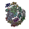
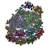
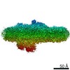
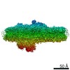
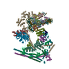
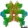
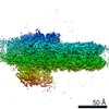
| Title | Structure of PSI-LHCI | ||||||||||||||||||||||||||||||||||||||||||||||||
 Components Components |
| ||||||||||||||||||||||||||||||||||||||||||||||||
 Keywords Keywords | PLANT PROTEIN / PSI-LHCI | ||||||||||||||||||||||||||||||||||||||||||||||||
| Function / homology |  Function and homology information Function and homology informationplastid thylakoid membrane / photosynthesis, light harvesting / chloroplast thylakoid lumen / photosystem I reaction center / photosystem I / photosynthetic electron transport in photosystem I / photosystem I / photosystem II / chlorophyll binding / chloroplast thylakoid membrane ...plastid thylakoid membrane / photosynthesis, light harvesting / chloroplast thylakoid lumen / photosystem I reaction center / photosystem I / photosynthetic electron transport in photosystem I / photosystem I / photosystem II / chlorophyll binding / chloroplast thylakoid membrane / photosynthesis / chloroplast / 4 iron, 4 sulfur cluster binding / electron transfer activity / oxidoreductase activity / protein domain specific binding / metal ion binding Similarity search - Function | ||||||||||||||||||||||||||||||||||||||||||||||||
| Biological species |  Bryopsis corticulans (plant) Bryopsis corticulans (plant) | ||||||||||||||||||||||||||||||||||||||||||||||||
| Method | ELECTRON MICROSCOPY / single particle reconstruction / cryo EM / Resolution: 3.49 Å | ||||||||||||||||||||||||||||||||||||||||||||||||
 Authors Authors | Xiong, P. / Xiaochun, Q. | ||||||||||||||||||||||||||||||||||||||||||||||||
 Citation Citation | Journal: Nat Plants / Year: 2019 Title: Structure of a green algal photosystem I in complex with a large number of light-harvesting complex I subunits. Authors: Xiaochun Qin / Xiong Pi / Wenda Wang / Guangye Han / Lixia Zhu / Mingmei Liu / Linpeng Cheng / Jian-Ren Shen / Tingyun Kuang / Sen-Fang Sui /   Abstract: Photosystem I (PSI) is a highly efficient natural light-energy converter, and has diverse light-harvesting antennas associated with its core in different photosynthetic organisms. In green algae, an ...Photosystem I (PSI) is a highly efficient natural light-energy converter, and has diverse light-harvesting antennas associated with its core in different photosynthetic organisms. In green algae, an extremely large light-harvesting complex I (LHCI) captures and transfers energy to the PSI core. Here, we report the structure of PSI-LHCI from a green alga Bryopsis corticulans at 3.49 Å resolution, obtained by single-particle cryo-electron microscopy, which revealed 13 core subunits including subunits characteristic of both prokaryotes and eukaryotes, and 10 light-harvesting complex a (Lhca) antennas that form a double semi-ring and an additional Lhca dimer, including a novel four-transmembrane-helix Lhca. In total, 244 chlorophylls were identified, some of which were located at key positions for the fast energy transfer. These results provide a firm structural basis for unravelling the mechanisms of light-energy harvesting, transfer and quenching in the green algal PSI-LHCI, and important clues as to how PSI-LHCI has changed during evolution. | ||||||||||||||||||||||||||||||||||||||||||||||||
| History |
|
- Structure visualization
| Movie |
Movie viewer |
|---|---|
| Structure viewer | Molecule: MolmilJmol/JSmol |
- Downloads & links
-Download
| PDBx/mmCIF format | 6igz.cif.gz | 1.2 MB | Display | PDBx/mmCIF format |
|---|---|---|---|---|
| PDB format | pdb6igz.ent.gz | 1.1 MB | Display | PDB format |
| PDBx/mmJSON format | 6igz.json.gz | Tree view | PDBx/mmJSON format | |
| Others |  Other downloads Other downloads |
-Validation report
| Arichive directory | https://data.pdbj.org/pub/pdb/validation_reports/ig/6igzftp://data.pdbj.org/pub/pdb/validation_reports/ig/6igz | HTTPS FTP |
|---|
-Related structure data
| Related structure data | 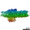 9670MC M: map data used to model this data C: citing same article ( |
|---|---|
| Similar structure data |
-Links
 PDBj
PDBj


- Assembly
| Deposited unit | 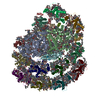
|
|---|---|
| 1 |
|
-Components
-Protein , 18 types, 20 molecules ABCDEFGHKL1523486790
| #1: Protein | Mass: 83412.336 Da / Num. of mol.: 1 / Source method: isolated from a natural source / Source: (natural) Bryopsis corticulans (plant) / References: UniProt: A0A4V8GZZ2*PLUS | ||||||||||||||
|---|---|---|---|---|---|---|---|---|---|---|---|---|---|---|---|
| #2: Protein | Mass: 82164.461 Da / Num. of mol.: 1 / Source method: isolated from a natural source / Source: (natural) Bryopsis corticulans (plant) / References: UniProt: A0A4V8H003*PLUS | ||||||||||||||
| #3: Protein | Mass: 8880.307 Da / Num. of mol.: 1 / Source method: isolated from a natural source / Source: (natural) Bryopsis corticulans (plant) / References: UniProt: A0A4V8H005*PLUS | ||||||||||||||
| #4: Protein | Mass: 21733.045 Da / Num. of mol.: 1 / Source method: isolated from a natural source / Source: (natural) Bryopsis corticulans (plant) / References: UniProt: A0A4V8H006*PLUS | ||||||||||||||
| #5: Protein | Mass: 10096.458 Da / Num. of mol.: 1 / Source method: isolated from a natural source / Source: (natural) Bryopsis corticulans (plant) / References: UniProt: A0A4V8H007*PLUS | ||||||||||||||
| #6: Protein | Mass: 25129.158 Da / Num. of mol.: 1 / Source method: isolated from a natural source / Source: (natural) Bryopsis corticulans (plant) / References: UniProt: A0A4V8H008*PLUS | ||||||||||||||
| #7: Protein | Mass: 18226.600 Da / Num. of mol.: 1 / Source method: isolated from a natural source / Source: (natural) Bryopsis corticulans (plant) | ||||||||||||||
| #8: Protein | Mass: 14400.360 Da / Num. of mol.: 1 / Source method: isolated from a natural source / Source: (natural) Bryopsis corticulans (plant) / References: UniProt: A0A4V8H009*PLUS | ||||||||||||||
| #11: Protein | Mass: 12487.672 Da / Num. of mol.: 1 / Source method: isolated from a natural source / Source: (natural) Bryopsis corticulans (plant) / References: UniProt: A0A4V8GZZ4*PLUS | ||||||||||||||
| #12: Protein | Mass: 21338.602 Da / Num. of mol.: 1 / Source method: isolated from a natural source / Source: (natural) Bryopsis corticulans (plant) / References: UniProt: A0A4V8GZZ5*PLUS | ||||||||||||||
| #13: Protein | Mass: 24458.012 Da / Num. of mol.: 2 / Source method: isolated from a natural source / Source: (natural) Bryopsis corticulans (plant) / References: UniProt: A0A4V8GZZ6*PLUS#14: Protein | | Mass: 27808.264 Da / Num. of mol.: 1 / Source method: isolated from a natural source / Source: (natural) Bryopsis corticulans (plant) / References: UniProt: A0A4V8GZZ7*PLUS#15: Protein | | Mass: 30409.689 Da / Num. of mol.: 1 / Source method: isolated from a natural source / Source: (natural) Bryopsis corticulans (plant) / References: UniProt: A0A4V8GZZ8*PLUS#16: Protein | Mass: 26575.912 Da / Num. of mol.: 2 / Source method: isolated from a natural source / Source: (natural) Bryopsis corticulans (plant) / References: UniProt: A0A4V8GZZ9*PLUS#17: Protein | | Mass: 29300.680 Da / Num. of mol.: 1 / Source method: isolated from a natural source / Source: (natural) Bryopsis corticulans (plant) / References: UniProt: A0A4V8H000*PLUS#18: Protein | | Mass: 28621.643 Da / Num. of mol.: 1 / Source method: isolated from a natural source / Source: (natural) Bryopsis corticulans (plant) / References: UniProt: A0A4V8H001*PLUS#19: Protein | | Mass: 24152.645 Da / Num. of mol.: 1 / Source method: isolated from a natural source / Source: (natural) Bryopsis corticulans (plant) / References: UniProt: A0A4V8H002*PLUS#20: Protein | | Mass: 26910.092 Da / Num. of mol.: 1 / Source method: isolated from a natural source / Source: (natural) Bryopsis corticulans (plant) / References: UniProt: A0A4V8H004*PLUS |
-Protein/peptide , 3 types, 3 molecules IJM
| #9: Protein/peptide | Mass: 3859.615 Da / Num. of mol.: 1 / Source method: isolated from a natural source / Source: (natural) Bryopsis corticulans (plant) / References: UniProt: A0A4V8H010*PLUS |
|---|---|
| #10: Protein/peptide | Mass: 4744.679 Da / Num. of mol.: 1 / Source method: isolated from a natural source / Source: (natural) Bryopsis corticulans (plant) / References: UniProt: A0A4V8GZZ3*PLUS |
| #21: Protein/peptide | Mass: 3505.114 Da / Num. of mol.: 1 / Source method: isolated from a natural source / Source: (natural) Bryopsis corticulans (plant) / References: UniProt: A0A0D6E2L8 |
-Sugars , 2 types, 3 molecules 

| #26: Sugar |  Type: D-saccharide / Mass: 294.408 Da / Num. of mol.: 2 / Source method: obtained synthetically / Formula: C13H26O5S / Comment: detergent*YM Type: D-saccharide / Mass: 294.408 Da / Num. of mol.: 2 / Source method: obtained synthetically / Formula: C13H26O5S / Comment: detergent*YM#28: Sugar | ChemComp-DGD / |  Type: saccharide / Mass: 949.299 Da / Num. of mol.: 1 / Source method: obtained synthetically / Formula: C51H96O15 Type: saccharide / Mass: 949.299 Da / Num. of mol.: 1 / Source method: obtained synthetically / Formula: C51H96O15 |
|---|
-Non-polymers , 8 types, 317 molecules 


| #22: Chemical | ChemComp-CLA /  Mass: 893.489 Da / Num. of mol.: 218 / Source method: obtained synthetically / Formula: C55H72MgN4O5 Mass: 893.489 Da / Num. of mol.: 218 / Source method: obtained synthetically / Formula: C55H72MgN4O5#23: Chemical |  Mass: 450.696 Da / Num. of mol.: 2 / Source method: obtained synthetically / Formula: C31H46O2 Mass: 450.696 Da / Num. of mol.: 2 / Source method: obtained synthetically / Formula: C31H46O2#24: Chemical | ChemComp-LHG /  Mass: 722.970 Da / Num. of mol.: 11 / Source method: obtained synthetically / Formula: C38H75O10P / Comment: phospholipid*YM Mass: 722.970 Da / Num. of mol.: 11 / Source method: obtained synthetically / Formula: C38H75O10P / Comment: phospholipid*YM#25: Chemical | ChemComp-8CT / (  Mass: 536.873 Da / Num. of mol.: 34 / Source method: obtained synthetically / Formula: C40H56 Mass: 536.873 Da / Num. of mol.: 34 / Source method: obtained synthetically / Formula: C40H56#27: Chemical |  Mass: 351.640 Da / Num. of mol.: 3 / Source method: obtained synthetically / Formula: Fe4S4 Mass: 351.640 Da / Num. of mol.: 3 / Source method: obtained synthetically / Formula: Fe4S4#29: Chemical | ChemComp-CHL /  Mass: 907.472 Da / Num. of mol.: 26 / Source method: obtained synthetically / Formula: C55H70MgN4O6 Mass: 907.472 Da / Num. of mol.: 26 / Source method: obtained synthetically / Formula: C55H70MgN4O6#30: Chemical | ChemComp-XAT / (  Mass: 600.870 Da / Num. of mol.: 20 / Source method: obtained synthetically / Formula: C40H56O4 Mass: 600.870 Da / Num. of mol.: 20 / Source method: obtained synthetically / Formula: C40H56O4#31: Chemical |  Mass: 787.158 Da / Num. of mol.: 3 / Source method: obtained synthetically / Formula: C45H86O10 Mass: 787.158 Da / Num. of mol.: 3 / Source method: obtained synthetically / Formula: C45H86O10 |
|---|
-Details
| Has protein modification | Y |
|---|
-Experimental details
-Experiment
| Experiment | Method: ELECTRON MICROSCOPY |
|---|---|
| EM experiment | Aggregation state: PARTICLE / 3D reconstruction method: single particle reconstruction |
- Sample preparation
| Component | Name: PSI-LHCI / Type: COMPLEX / Entity ID: #1-#21 / Source: NATURAL |
|---|---|
| Source (natural) | Organism: Bryopsis corticulans (plant) |
| Buffer solution | pH: 7.8 |
| Specimen | Conc.: 3 mg/ml / Embedding applied: NO / Shadowing applied: NO / Staining applied: NO / Vitrification applied: YES |
| Vitrification | Cryogen name: ETHANE |
- Electron microscopy imaging
| Experimental equipment |  Model: Titan Krios / Image courtesy: FEI Company |
|---|---|
| Microscopy | Model: FEI TITAN KRIOS |
| Electron gun | Electron source:  FIELD EMISSION GUN / Accelerating voltage: 300 kV / Illumination mode: FLOOD BEAM FIELD EMISSION GUN / Accelerating voltage: 300 kV / Illumination mode: FLOOD BEAM |
| Electron lens | Mode: BRIGHT FIELD |
| Image recording | Electron dose: 1.852 e/Å2 / Film or detector model: FEI FALCON II (4k x 4k) |
- Processing
| Software | Name: PHENIX / Version: 1.10.1_2155: / Classification: refinement | ||||||||||||||||||||||||
|---|---|---|---|---|---|---|---|---|---|---|---|---|---|---|---|---|---|---|---|---|---|---|---|---|---|
| EM software |
| ||||||||||||||||||||||||
| CTF correction | Type: NONE | ||||||||||||||||||||||||
| 3D reconstruction | Resolution: 3.49 Å / Resolution method: FSC 0.143 CUT-OFF / Num. of particles: 59525 / Symmetry type: POINT | ||||||||||||||||||||||||
| Refine LS restraints |
|
