 Movie
Movie Controller

[English] 日本語
 Yorodumi
- PDB-6ifi: Crystal Structure of the Apo form of CMP-N-acetylneuraminate Synt... -
+ Open data

- Basic information
| Entry | Database: PDB / ID: 6ifi | ||||||||||||
|---|---|---|---|---|---|---|---|---|---|---|---|---|---|
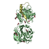
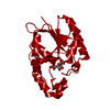
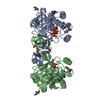
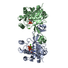
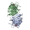
| Title | Crystal Structure of the Apo form of CMP-N-acetylneuraminate Synthetase from Vibrio cholerae | ||||||||||||
 Components Components | CMP-N-acetylneuraminate Synthetase | ||||||||||||
 Keywords Keywords | SUGAR BINDING PROTEIN / Bacterial / CMP-sialic acid synthetase / sialic acid / CTP | ||||||||||||
| Function / homology | Spore Coat Polysaccharide Biosynthesis Protein SpsA; Chain A / Spore Coat Polysaccharide Biosynthesis Protein SpsA; Chain A / Alpha-Beta Complex / Alpha Beta Function and homology information Function and homology information | ||||||||||||
| Biological species |   Vibrio cholerae (bacteria) Vibrio cholerae (bacteria) | ||||||||||||
| Method |  X-RAY DIFFRACTION / SYNCHROTRON / MOLECULAR REPLACEMENT / Resolution: 2.8 Å X-RAY DIFFRACTION / SYNCHROTRON / MOLECULAR REPLACEMENT / Resolution: 2.8 Å | ||||||||||||
 Authors Authors | Bose, S. / Subramanian, R. | ||||||||||||
| Funding support |  India, 3items India, 3items
| ||||||||||||
 Citation Citation | Journal: Acta Crystallogr D Struct Biol / Year: 2019 Title: Structural and functional characterization of CMP-N-acetylneuraminate synthetase from Vibrio cholerae. Authors: Bose, S. / Purkait, D. / Joseph, D. / Nayak, V. / Subramanian, R. | ||||||||||||
| History |
|
- Structure visualization
| Structure viewer | Molecule: MolmilJmol/JSmol |
|---|
- Downloads & links
-Download
| PDBx/mmCIF format | 6ifi.cif.gz | 99.7 KB | Display | PDBx/mmCIF format |
|---|---|---|---|---|
| PDB format | pdb6ifi.ent.gz | 73.7 KB | Display | PDB format |
| PDBx/mmJSON format | 6ifi.json.gz | Tree view | PDBx/mmJSON format | |
| Others |  Other downloads Other downloads |
-Validation report
| Arichive directory | https://data.pdbj.org/pub/pdb/validation_reports/if/6ififtp://data.pdbj.org/pub/pdb/validation_reports/if/6ifi | HTTPS FTP |
|---|
-Related structure data
| Related structure data |  6ifdC  1eziS S: Starting model for refinement C: citing same article ( |
|---|---|
| Similar structure data |


-Links
 PDBj
PDBj- Assembly
| Deposited unit | 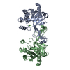
| ||||||||
|---|---|---|---|---|---|---|---|---|---|
| 1 |
| ||||||||
| Unit cell |
|
-Components
| #1: Protein | Mass: 28473.449 Da / Num. of mol.: 2 Source method: isolated from a genetically manipulated source Source: (gene. exp.) Vibrio cholerae (bacteria) / Production host: #2: Chemical |   Mass: 40.078 Da / Num. of mol.: 2 / Source method: obtained synthetically / Formula: Ca Mass: 40.078 Da / Num. of mol.: 2 / Source method: obtained synthetically / Formula: Ca#3: Water | ChemComp-HOH / |  Mass: 18.015 Da / Num. of mol.: 66 / Source method: isolated from a natural source / Formula: H2O Mass: 18.015 Da / Num. of mol.: 66 / Source method: isolated from a natural source / Formula: H2O |
|---|
-Experimental details
-Experiment
| Experiment | Method: X-RAY DIFFRACTION / Number of used crystals: 1 |
|---|
- Sample preparation
| Crystal | Density Matthews: 2.73 Å3/Da / Density % sol: 54.91 % |
|---|---|
| Crystal grow | Temperature: 291 K / Method: vapor diffusion, hanging drop / pH: 8 / Details: PEG 8000, calcium acetate, imidazole |
-Data collection
| Diffraction | Mean temperature: 100 K |
|---|---|
| Diffraction source | Source: SYNCHROTRON / Site: SOLEIL  / Beamline: PROXIMA 1 / Wavelength: 0.9786 Å / Beamline: PROXIMA 1 / Wavelength: 0.9786 Å |
| Detector | Type: DECTRIS PILATUS 6M / Detector: PIXEL / Date: Jun 28, 2016 |
| Radiation | Protocol: SINGLE WAVELENGTH / Monochromatic (M) / Laue (L): M / Scattering type: x-ray |
| Radiation wavelength | Wavelength: 0.9786 Å / Relative weight: 1 |
| Reflection | Resolution: 2.8→47.87 Å / Num. obs: 14951 / % possible obs: 99.9 % / Redundancy: 6.7 % / CC1/2: 0.99 / Rmerge(I) obs: 0.07 / Net I/σ(I): 16.7 |
| Reflection shell | Resolution: 2.8→2.95 Å / Rmerge(I) obs: 0.86 / Num. unique obs: 356 / CC1/2: 0.88 |
- Processing
| Software |
| ||||||||||||||||||||||||||||||||||||||||||||||||||||||||||||||||||||||||||||||||||||||||||||||||||||||||||||
|---|---|---|---|---|---|---|---|---|---|---|---|---|---|---|---|---|---|---|---|---|---|---|---|---|---|---|---|---|---|---|---|---|---|---|---|---|---|---|---|---|---|---|---|---|---|---|---|---|---|---|---|---|---|---|---|---|---|---|---|---|---|---|---|---|---|---|---|---|---|---|---|---|---|---|---|---|---|---|---|---|---|---|---|---|---|---|---|---|---|---|---|---|---|---|---|---|---|---|---|---|---|---|---|---|---|---|---|---|---|
| Refinement | Method to determine structure: MOLECULAR REPLACEMENT Starting model: 1EZI Resolution: 2.8→25.08 Å / Cor.coef. Fo:Fc: 0.924 / Cor.coef. Fo:Fc free: 0.905 / SU R Cruickshank DPI: 0.881 / Cross valid method: THROUGHOUT / σ(F): 0 / SU R Blow DPI: 0.749 / SU Rfree Blow DPI: 0.308 / SU Rfree Cruickshank DPI: 0.319
| ||||||||||||||||||||||||||||||||||||||||||||||||||||||||||||||||||||||||||||||||||||||||||||||||||||||||||||
| Displacement parameters | Biso max: 124.23 Å2 / Biso mean: 64.35 Å2 / Biso min: 23.05 Å2
| ||||||||||||||||||||||||||||||||||||||||||||||||||||||||||||||||||||||||||||||||||||||||||||||||||||||||||||
| Refine analyze | Luzzati coordinate error obs: 0.4 Å | ||||||||||||||||||||||||||||||||||||||||||||||||||||||||||||||||||||||||||||||||||||||||||||||||||||||||||||
| Refinement step | Cycle: final / Resolution: 2.8→25.08 Å
| ||||||||||||||||||||||||||||||||||||||||||||||||||||||||||||||||||||||||||||||||||||||||||||||||||||||||||||
| Refine LS restraints |
| ||||||||||||||||||||||||||||||||||||||||||||||||||||||||||||||||||||||||||||||||||||||||||||||||||||||||||||
| LS refinement shell | Resolution: 2.8→2.83 Å / Rfactor Rfree error: 0 / Total num. of bins used: 42
|
