 Movie
Movie Controller


+ Open data

- Basic information
| Entry | Database: PDB / ID: 6ifa | ||||||
|---|---|---|---|---|---|---|---|
| Title | Structure of beta-trefoil lectin from Entamoeba histolytica | ||||||
 Components Components | lectin | ||||||
 Keywords Keywords | SUGAR BINDING PROTEIN / beta-trefoil / lectin / Entamoeba histolytica | ||||||
| Function / homology | Ricin-type beta-trefoil lectin domain / Ricin-type beta-trefoil / Ricin B, lectin domain / Ricin B-like lectins / carbohydrate binding / ISOPROPYL ALCOHOL / Ricin-type beta-trefoil lectin domain containing protein Function and homology information Function and homology information | ||||||
| Biological species |  Entamoeba histolytica HM-1:IMSS (eukaryote) Entamoeba histolytica HM-1:IMSS (eukaryote) | ||||||
| Method |  X-RAY DIFFRACTION / SYNCHROTRON / MOLECULAR REPLACEMENT / Resolution: 1.9 Å X-RAY DIFFRACTION / SYNCHROTRON / MOLECULAR REPLACEMENT / Resolution: 1.9 Å | ||||||
 Authors Authors | Suguna, K. / Khan, F. | ||||||
 Citation Citation | Journal: Glycobiology / Year: 2020 Title: Crystal structures of a beta-trefoil lectin from Entamoeba histolytica in monomeric and a novel disulfide bond-mediated dimeric forms. Authors: Khan, F. / Kurre, D. / Suguna, K. | ||||||
| History |
|
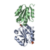
- Structure visualization
| Structure viewer | Molecule: MolmilJmol/JSmol |
|---|
- Downloads & links
-Download
| PDBx/mmCIF format | 6ifa.cif.gz | 113.2 KB | Display | PDBx/mmCIF format |
|---|---|---|---|---|
| PDB format | pdb6ifa.ent.gz | 71.6 KB | Display | PDB format |
| PDBx/mmJSON format | 6ifa.json.gz | Tree view | PDBx/mmJSON format | |
| Others |  Other downloads Other downloads |
-Validation report
| Summary document | 6ifa_validation.pdf.gz | 463.2 KB | Display | wwPDB validaton report |
|---|---|---|---|---|
| Full document | 6ifa_full_validation.pdf.gz | 464.6 KB | Display | |
| Data in XML | 6ifa_validation.xml.gz | 17.8 KB | Display | |
| Data in CIF | 6ifa_validation.cif.gz | 24.2 KB | Display | |
| Arichive directory | https://data.pdbj.org/pub/pdb/validation_reports/if/6ifaftp://data.pdbj.org/pub/pdb/validation_reports/if/6ifa | HTTPS FTP |
-Related structure data


-Links
 PDBj
PDBj


- Assembly
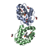
| Deposited unit | 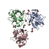
| ||||||||||||
|---|---|---|---|---|---|---|---|---|---|---|---|---|---|
| 1 | 
| ||||||||||||
| 2 | 
| ||||||||||||
| Unit cell |
|
-Components
| #1: Protein | Mass: 14677.430 Da / Num. of mol.: 3 Source method: isolated from a genetically manipulated source Source: (gene. exp.) Entamoeba histolytica HM-1:IMSS (eukaryote)Production host:  References: UniProt: N9TFI9*PLUS #2: Chemical | ChemComp-GOL /   Mass: 92.094 Da / Num. of mol.: 5 / Source method: obtained synthetically / Formula: C3H8O3 Mass: 92.094 Da / Num. of mol.: 5 / Source method: obtained synthetically / Formula: C3H8O3#3: Chemical | ChemComp-IPA /   Mass: 60.095 Da / Num. of mol.: 9 / Source method: obtained synthetically / Formula: C3H8O Mass: 60.095 Da / Num. of mol.: 9 / Source method: obtained synthetically / Formula: C3H8O#4: Water | ChemComp-HOH / |  Mass: 18.015 Da / Num. of mol.: 171 / Source method: isolated from a natural source / Formula: H2O Mass: 18.015 Da / Num. of mol.: 171 / Source method: isolated from a natural source / Formula: H2OHas protein modification | Y | |
|---|
-Experimental details
-Experiment
| Experiment | Method: X-RAY DIFFRACTION / Number of used crystals: 1 |
|---|
- Sample preparation
| Crystal | Density Matthews: 2.77 Å3/Da / Density % sol: 55.52 % |
|---|---|
| Crystal grow | Temperature: 289.15 K / Method: vapor diffusion, hanging drop Details: 20% iso-propanol, 0.1M MES monohydrate, pH 6.0, 20% PEG monomethyl ether 2000 |
-Data collection
| Diffraction | Mean temperature: 100 K / Serial crystal experiment: N |
|---|---|
| Diffraction source | Source: SYNCHROTRON / Site: ESRF  / Beamline: MASSIF-1 / Wavelength: 0.966 Å / Beamline: MASSIF-1 / Wavelength: 0.966 Å |
| Detector | Type: DECTRIS PILATUS3 2M / Detector: PIXEL / Date: Jul 14, 2018 |
| Radiation | Protocol: SINGLE WAVELENGTH / Monochromatic (M) / Laue (L): M / Scattering type: x-ray |
| Radiation wavelength | Wavelength: 0.966 Å / Relative weight: 1 |
| Reflection | Resolution: 1.9→46 Å / Num. obs: 38368 / % possible obs: 99.9 % / Redundancy: 13.23 % / Biso Wilson estimate: 27.15 Å2 / CC1/2: 0.99 / Net I/σ(I): 13.88 |
| Reflection shell | Resolution: 1.9→2.01 Å / Redundancy: 12.43 % / Mean I/σ(I) obs: 1.75 / Num. unique obs: 6102 / CC1/2: 0.84 / % possible all: 99.8 |
- Processing
| Software |
| ||||||||||||||||||||||||
|---|---|---|---|---|---|---|---|---|---|---|---|---|---|---|---|---|---|---|---|---|---|---|---|---|---|
| Refinement | Method to determine structure: MOLECULAR REPLACEMENT / Resolution: 1.9→46 Å / SU ML: 0.2458 / Cross valid method: FREE R-VALUE / Phase error: 25.6638
| ||||||||||||||||||||||||
| Solvent computation | Shrinkage radii: 0.9 Å / VDW probe radii: 1.11 Å | ||||||||||||||||||||||||
| Displacement parameters | Biso mean: 31.51 Å2 | ||||||||||||||||||||||||
| Refinement step | Cycle: LAST / Resolution: 1.9→46 Å
| ||||||||||||||||||||||||
| Refine LS restraints |
| ||||||||||||||||||||||||
| LS refinement shell | Resolution: 1.9→1.95 Å
|
