 Movie
Movie Controller

[English] 日本語
 Yorodumi
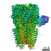
- PDB-6i53: Cryo-EM structure of the human synaptic alpha1-beta3-gamma2 GABAA... -
+ Open data

- Basic information
| Entry | Database: PDB / ID: 6i53 | ||||||||||||||||||||||||
|---|---|---|---|---|---|---|---|---|---|---|---|---|---|---|---|---|---|---|---|---|---|---|---|---|---|
| Title | Cryo-EM structure of the human synaptic alpha1-beta3-gamma2 GABAA receptor in complex with Megabody38 in a lipid nanodisc | ||||||||||||||||||||||||
 Components Components |
| ||||||||||||||||||||||||
 Keywords Keywords | TRANSPORT PROTEIN / Ligand gated ion channel Cys-Loop receptor GABA-A receptor | ||||||||||||||||||||||||
| Function / homology |  Function and homology information Function and homology informationbenzodiazepine receptor activity / circadian sleep/wake cycle, REM sleep / reproductive behavior / GABA receptor complex / hard palate development / cellular response to histamine / GABA receptor activation / inner ear receptor cell development / innervation / GABA-gated chloride ion channel activity ...benzodiazepine receptor activity / circadian sleep/wake cycle, REM sleep / reproductive behavior / GABA receptor complex / hard palate development / cellular response to histamine / GABA receptor activation / inner ear receptor cell development / innervation / GABA-gated chloride ion channel activity / GABA-A receptor complex / inhibitory synapse assembly / GABA-A receptor activity / response to anesthetic / inhibitory postsynaptic potential / postsynaptic specialization membrane / roof of mouth development / gamma-aminobutyric acid signaling pathway / synaptic transmission, GABAergic / adult behavior / exploration behavior / motor behavior / cellular response to zinc ion / chloride channel activity / Signaling by ERBB4 / cochlea development / social behavior / chloride channel complex / post-embryonic development / cerebellum development / dendrite membrane / cytoplasmic vesicle membrane / chloride transmembrane transport / learning / transmitter-gated monoatomic ion channel activity involved in regulation of postsynaptic membrane potential / GABA-ergic synapse / memory / dendritic spine / postsynaptic membrane / postsynapse / response to xenobiotic stimulus / axon / cell surface / signal transduction / identical protein binding / plasma membrane Similarity search - Function | ||||||||||||||||||||||||
| Biological species |  Homo sapiens (human) Homo sapiens (human) | ||||||||||||||||||||||||
| Method | ELECTRON MICROSCOPY / single particle reconstruction / cryo EM / Resolution: 3.2 Å | ||||||||||||||||||||||||
 Authors Authors | Laverty, D. / Desai, R. / Uchanski, T. / Masiulis, S. / Wojciech, J.S. / Malinauskas, T. / Zivanov, J. / Pardon, E. / Steyaert, J. / Miller, K.W. / Aricescu, A.R. | ||||||||||||||||||||||||
| Funding support |  United Kingdom, United Kingdom,  Switzerland, Switzerland,  United States, 7items United States, 7items
| ||||||||||||||||||||||||
 Citation Citation | Journal: Nature / Year: 2019 Title: Cryo-EM structure of the human α1β3γ2 GABA receptor in a lipid bilayer. Authors: Duncan Laverty / Rooma Desai / Tomasz Uchański / Simonas Masiulis / Wojciech J Stec / Tomas Malinauskas / Jasenko Zivanov / Els Pardon / Jan Steyaert / Keith W Miller / A Radu Aricescu /  Abstract: Type A γ-aminobutyric acid (GABA) receptors are pentameric ligand-gated ion channels and the main drivers of fast inhibitory neurotransmission in the vertebrate nervous system. Their dysfunction is ...Type A γ-aminobutyric acid (GABA) receptors are pentameric ligand-gated ion channels and the main drivers of fast inhibitory neurotransmission in the vertebrate nervous system. Their dysfunction is implicated in a range of neurological disorders, including depression, epilepsy and schizophrenia. Among the numerous assemblies that are theoretically possible, the most prevalent in the brain are the α1β2/3γ2 GABA receptors. The β3 subunit has an important role in maintaining inhibitory tone, and the expression of this subunit alone is sufficient to rescue inhibitory synaptic transmission in β1-β3 triple knockout neurons. So far, efforts to generate accurate structural models for heteromeric GABA receptors have been hampered by the use of engineered receptors and the presence of detergents. Notably, some recent cryo-electron microscopy reconstructions have reported 'collapsed' conformations; however, these disagree with the structure of the prototypical pentameric ligand-gated ion channel the Torpedo nicotinic acetylcholine receptor, the large body of structural work on homologous homopentameric receptor variants and the logic of an ion-channel architecture. Here we present a high-resolution cryo-electron microscopy structure of the full-length human α1β3γ2L-a major synaptic GABA receptor isoform-that is functionally reconstituted in lipid nanodiscs. The receptor is bound to a positive allosteric modulator 'megabody' and is in a desensitized conformation. Each GABA receptor pentamer contains two phosphatidylinositol-4,5-bisphosphate molecules, the head groups of which occupy positively charged pockets in the intracellular juxtamembrane regions of α1 subunits. Beyond this level, the intracellular M3-M4 loops are largely disordered, possibly because interacting post-synaptic proteins are not present. This structure illustrates the molecular principles of heteromeric GABA receptor organization and provides a reference framework for future mechanistic investigations of GABAergic signalling and pharmacology. | ||||||||||||||||||||||||
| History |
|
- Structure visualization
| Movie |
Movie viewer |
|---|---|
| Structure viewer | Molecule: MolmilJmol/JSmol |
- Downloads & links
-Download
| PDBx/mmCIF format | 6i53.cif.gz | 366.9 KB | Display | PDBx/mmCIF format |
|---|---|---|---|---|
| PDB format | pdb6i53.ent.gz | 284.3 KB | Display | PDB format |
| PDBx/mmJSON format | 6i53.json.gz | Tree view | PDBx/mmJSON format | |
| Others |  Other downloads Other downloads |
-Validation report
| Arichive directory | https://data.pdbj.org/pub/pdb/validation_reports/i5/6i53ftp://data.pdbj.org/pub/pdb/validation_reports/i5/6i53 | HTTPS FTP |
|---|
-Related structure data
| Related structure data | 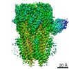 4411MC M: map data used to model this data C: citing same article ( |
|---|---|
| Similar structure data |


-Links
 PDBj
PDBj


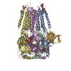
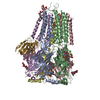
- Assembly
| Deposited unit | 
|
|---|---|
| 1 |
|
-Components
-Gamma-aminobutyric acid receptor subunit ... , 3 types, 5 molecules EBADC
| #1: Protein | Mass: 54444.578 Da / Num. of mol.: 2 Source method: isolated from a genetically manipulated source Source: (gene. exp.) Homo sapiens (human) / Gene: GABRB3 / Cell line (production host): HEK293 / Production host: Homo sapiens (human) / References: UniProt: P28472#2: Protein | Mass: 52916.602 Da / Num. of mol.: 2 Source method: isolated from a genetically manipulated source Source: (gene. exp.) Homo sapiens (human) / Gene: GABRA1 / Cell line (production host): HEK293 / Production host: Homo sapiens (human) / References: UniProt: P14867#3: Protein | | Mass: 56922.055 Da / Num. of mol.: 1 Source method: isolated from a genetically manipulated source Source: (gene. exp.) Homo sapiens (human) / Gene: GABRG2 / Cell line (production host): HEK293 / Production host: Homo sapiens (human) / References: UniProt: P18507 |
|---|
-Antibody , 1 types, 1 molecules G
| #4: Antibody | Mass: 13462.909 Da / Num. of mol.: 1 Source method: isolated from a genetically manipulated source Source: (gene. exp.)  |
|---|
-Sugars , 4 types, 7 molecules
| #5: Polysaccharide | Source method: isolated from a genetically manipulated source #6: Polysaccharide | Source method: isolated from a genetically manipulated source #7: Polysaccharide | alpha-D-mannopyranose-(1-3)-[alpha-D-mannopyranose-(1-6)]alpha-D-mannopyranose-(1-6)-[alpha-D- ...alpha-D-mannopyranose-(1-3)-[alpha-D-mannopyranose-(1-6)]alpha-D-mannopyranose-(1-6)-[alpha-D-mannopyranose-(1-3)]beta-D-mannopyranose-(1-4)-2-acetamido-2-deoxy-beta-D-glucopyranose-(1-4)-2-acetamido-2-deoxy-beta-D-glucopyranose | Source method: isolated from a genetically manipulated source #8: Polysaccharide | alpha-D-mannopyranose-(1-3)-alpha-D-mannopyranose-(1-6)-[alpha-D-mannopyranose-(1-3)]beta-D- ...alpha-D-mannopyranose-(1-3)-alpha-D-mannopyranose-(1-6)-[alpha-D-mannopyranose-(1-3)]beta-D-mannopyranose-(1-4)-2-acetamido-2-deoxy-beta-D-glucopyranose-(1-4)-2-acetamido-2-deoxy-beta-D-glucopyranose | Source method: isolated from a genetically manipulated source |
|---|
-Non-polymers , 2 types, 5 molecules 

| #9: Chemical |  Mass: 760.076 Da / Num. of mol.: 3 / Source method: obtained synthetically / Formula: C42H82NO8P / Comment: phospholipid*YM Mass: 760.076 Da / Num. of mol.: 3 / Source method: obtained synthetically / Formula: C42H82NO8P / Comment: phospholipid*YM#10: Chemical |  Mass: 746.566 Da / Num. of mol.: 2 Mass: 746.566 Da / Num. of mol.: 2Source method: isolated from a genetically manipulated source Formula: C25H49O19P3 |
|---|
-Details
| Has protein modification | Y |
|---|
-Experimental details
-Experiment
| Experiment | Method: ELECTRON MICROSCOPY |
|---|---|
| EM experiment | Aggregation state: PARTICLE / 3D reconstruction method: single particle reconstruction |
- Sample preparation
| Component |
| ||||||||||||||||||||||||
|---|---|---|---|---|---|---|---|---|---|---|---|---|---|---|---|---|---|---|---|---|---|---|---|---|---|
| Molecular weight | Value: 0.4 MDa / Experimental value: NO | ||||||||||||||||||||||||
| Source (natural) |
| ||||||||||||||||||||||||
| Source (recombinant) |
| ||||||||||||||||||||||||
| Buffer solution | pH: 7.2 | ||||||||||||||||||||||||
| Buffer component |
| ||||||||||||||||||||||||
| Specimen | Conc.: 0.5 mg/ml / Embedding applied: NO / Shadowing applied: NO / Staining applied: NO / Vitrification applied: YES Details: The purified receptor-nanodisc complex was mixed with Mb38 prior to grid preparation. | ||||||||||||||||||||||||
| Specimen support | Grid material: GOLD / Grid type: Quantifoil, UltrAuFoil | ||||||||||||||||||||||||
| Vitrification | Instrument: FEI VITROBOT MARK IV / Cryogen name: ETHANE / Humidity: 100 % / Chamber temperature: 287.15 K |
- Electron microscopy imaging
| Experimental equipment |  Model: Titan Krios / Image courtesy: FEI Company |
|---|---|
| Microscopy | Model: FEI TITAN KRIOS |
| Electron gun | Electron source:  FIELD EMISSION GUN / Accelerating voltage: 300 kV / Illumination mode: FLOOD BEAM FIELD EMISSION GUN / Accelerating voltage: 300 kV / Illumination mode: FLOOD BEAM |
| Electron lens | Mode: BRIGHT FIELD / Nominal magnification: 75000 X / Calibrated magnification: 75000 X / Nominal defocus max: 700 nm / Nominal defocus min: 500 nm / Calibrated defocus min: 500 nm / Calibrated defocus max: 700 nm / Cs: 2.7 mm / C2 aperture diameter: 50 µm |
| Specimen holder | Specimen holder model: FEI TITAN KRIOS AUTOGRID HOLDER |
| Image recording | Average exposure time: 60 sec. / Electron dose: 30.84 e/Å2 / Detector mode: COUNTING / Film or detector model: FEI FALCON III (4k x 4k) / Num. of grids imaged: 1 / Num. of real images: 784 |
| EM imaging optics | Phase plate: VOLTA PHASE PLATE |
- Processing
| Software | Name: PHENIX / Version: 1.14rc2_3191: / Classification: refinement | ||||||||||||||||||||||||||||
|---|---|---|---|---|---|---|---|---|---|---|---|---|---|---|---|---|---|---|---|---|---|---|---|---|---|---|---|---|---|
| EM software |
| ||||||||||||||||||||||||||||
| CTF correction | Details: CTF parameters were estimated using GCTF and CTF correction performed in RELION (full phase and amplitude correction). Type: PHASE FLIPPING AND AMPLITUDE CORRECTION | ||||||||||||||||||||||||||||
| Symmetry | Point symmetry: C1 (asymmetric) | ||||||||||||||||||||||||||||
| 3D reconstruction | Resolution: 3.2 Å / Resolution method: FSC 0.143 CUT-OFF / Num. of particles: 55449 / Algorithm: FOURIER SPACE / Num. of class averages: 1 / Symmetry type: POINT | ||||||||||||||||||||||||||||
| Atomic model building | Protocol: AB INITIO MODEL / Space: REAL | ||||||||||||||||||||||||||||
| Refinement | Stereochemistry target values: CDL v1.2 | ||||||||||||||||||||||||||||
| Refine LS restraints |
|
