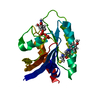
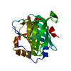
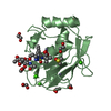
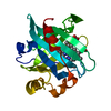
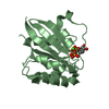
Entry Database : PDB / ID : 6i42Title Structure of the alpha-Synuclein PreNAC/Cyclophilin A-complex Alpha-synuclein Peptidyl-prolyl cis-trans isomerase A Keywords / / / / Function / homology Function Domain/homology Component
/ / / / / / / / / / / / / / / / / / / / / / / / / / / / / / / / / / / / / / / / / / / / / / / / / / / / / / / / / / / / / / / / / / / / / / / / / / / / / / / / / / / / / / / / / / / / / / / / / / / / / / / / / / / / / / / / / / / / / / / / / / / / / / / / / / Biological species Homo sapiens (human)Method / / / / Resolution : 1.38 Å Authors Favretto, F. / Zweckstetter, M. / Becker, S. Journal : Angew.Chem.Int.Ed.Engl. / Year : 2020Title : The Molecular Basis of the Interaction of Cyclophilin A with alpha-Synuclein.Authors : Favretto, F. / Baker, J.D. / Strohaker, T. / Andreas, L.B. / Blair, L.J. / Becker, S. / Zweckstetter, M. History Deposition Nov 8, 2018 Deposition site / Processing site Revision 1.0 Feb 19, 2020 Provider / Type Revision 1.1 Apr 1, 2020 Group / Category / citation_authorItem _citation.journal_volume / _citation.page_first ... _citation.journal_volume / _citation.page_first / _citation.page_last / _citation.year / _citation_author.identifier_ORCID Revision 1.2 Jan 24, 2024 Group / Database references / Refinement descriptionCategory chem_comp_atom / chem_comp_bond ... chem_comp_atom / chem_comp_bond / database_2 / pdbx_initial_refinement_model Item / _database_2.pdbx_database_accession
Show all Show less
 Movie
Movie Controller


 Open data
 Basic information
 Components
 Keywords
 Function and homology information
 Homo sapiens (human)
 X-RAY DIFFRACTION /
  Authors
 Citation
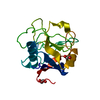
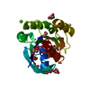
 Structure visualization
 Downloads & links
 Other downloads


 PDBj
PDBj


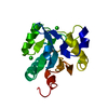
 Assembly
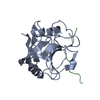

 Mass: 18.015 Da / Num. of mol.: 248 / Source method: isolated from a natural source / Formula: H2O
 Sample preparation
 / Beamline: X10SA / Wavelength: 1 Å
 Processing