 Movie
Movie Controller

[English] 日本語
 Yorodumi
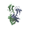
- PDB-6hwp: Structure of A3_bGFPD, an artificial bi-domain protein based on t... -
+ Open data

- Basic information
| Entry | Database: PDB / ID: 6hwp | ||||||
|---|---|---|---|---|---|---|---|
| Title | Structure of A3_bGFPD, an artificial bi-domain protein based on two different alphaRep domains : A3 and a GFP binding domain (bGFPD) | ||||||
 Components Components | A3_bGFPD | ||||||
 Keywords Keywords | BIOSYNTHETIC PROTEIN / alphaRep / artificial protein / chimera / bidomain | ||||||
| Function / homology | MALONATE ION Function and homology information Function and homology information | ||||||
| Biological species | synthetic construct (others) | ||||||
| Method |  X-RAY DIFFRACTION / SYNCHROTRON / MOLECULAR REPLACEMENT / molecular replacement / Resolution: 2.547 Å X-RAY DIFFRACTION / SYNCHROTRON / MOLECULAR REPLACEMENT / molecular replacement / Resolution: 2.547 Å | ||||||
 Authors Authors | Li de la Sierra-Gallay, I. / Leger, C. | ||||||
| Funding support |  France, 1items France, 1items
| ||||||
 Citation Citation | Journal: Sci Rep / Year: 2019 Title: Ligand-induced conformational switch in an artificial bidomain protein scaffold. Authors: Leger, C. / Di Meo, T. / Aumont-Nicaise, M. / Velours, C. / Durand, D. / Li de la Sierra-Gallay, I. / van Tilbeurgh, H. / Hildebrandt, N. / Desmadril, M. / Urvoas, A. / Valerio-Lepiniec, M. / Minard, P. #1: Journal: To Be PublishedTitle: Bi-domain artificial protein alphaRep display inducible cooperativity Authors: Leger, C. / Li de la Sierra-Gallay, I. | ||||||
| History |
|
- Structure visualization
| Structure viewer | Molecule: MolmilJmol/JSmol |
|---|
- Downloads & links
-Download
| PDBx/mmCIF format | 6hwp.cif.gz | 82.5 KB | Display | PDBx/mmCIF format |
|---|---|---|---|---|
| PDB format | pdb6hwp.ent.gz | 61.4 KB | Display | PDB format |
| PDBx/mmJSON format | 6hwp.json.gz | Tree view | PDBx/mmJSON format | |
| Others |  Other downloads Other downloads |
-Validation report
| Summary document | 6hwp_validation.pdf.gz | 443.9 KB | Display | wwPDB validaton report |
|---|---|---|---|---|
| Full document | 6hwp_full_validation.pdf.gz | 447.1 KB | Display | |
| Data in XML | 6hwp_validation.xml.gz | 14 KB | Display | |
| Data in CIF | 6hwp_validation.cif.gz | 18.2 KB | Display | |
| Arichive directory | https://data.pdbj.org/pub/pdb/validation_reports/hw/6hwpftp://data.pdbj.org/pub/pdb/validation_reports/hw/6hwp | HTTPS FTP |
-Related structure data
| Related structure data |  6fsqC  6ft5C  3ltjS C: citing same article ( S: Starting model for refinement |
|---|---|
| Similar structure data |


-Links
 PDBj
PDBj- Assembly
| Deposited unit | 
| ||||||||
|---|---|---|---|---|---|---|---|---|---|
| 1 |
| ||||||||
| Unit cell |
|
-Components
| #1: Protein | Mass: 44460.203 Da / Num. of mol.: 1 Source method: isolated from a genetically manipulated source Source: (gene. exp.) synthetic construct (others) / Production host:  |
|---|---|
| #2: Chemical | ChemComp-MLI /   Mass: 102.046 Da / Num. of mol.: 1 / Source method: obtained synthetically / Formula: C3H2O4 Mass: 102.046 Da / Num. of mol.: 1 / Source method: obtained synthetically / Formula: C3H2O4 |
| #3: Chemical | ChemComp-NA /   Mass: 22.990 Da / Num. of mol.: 1 / Source method: obtained synthetically / Formula: Na Mass: 22.990 Da / Num. of mol.: 1 / Source method: obtained synthetically / Formula: Na |
-Experimental details
-Experiment
| Experiment | Method: X-RAY DIFFRACTION / Number of used crystals: 1 |
|---|
- Sample preparation
| Crystal | Density Matthews: 2.4 Å3/Da / Density % sol: 48.83 % |
|---|---|
| Crystal grow | Temperature: 293 K / Method: vapor diffusion, sitting drop / pH: 6.5 / Details: 1.6M Tri-sodium citrate |
-Data collection
| Diffraction | Mean temperature: 100 K / Serial crystal experiment: N | ||||||||||||||||||||||||||||||||||||||||||||||||||||||||||||||||||||||||||||||||
|---|---|---|---|---|---|---|---|---|---|---|---|---|---|---|---|---|---|---|---|---|---|---|---|---|---|---|---|---|---|---|---|---|---|---|---|---|---|---|---|---|---|---|---|---|---|---|---|---|---|---|---|---|---|---|---|---|---|---|---|---|---|---|---|---|---|---|---|---|---|---|---|---|---|---|---|---|---|---|---|---|---|
| Diffraction source | Source: SYNCHROTRON / Site: ESRF / Beamline: ID29 / Wavelength: 0.97 Å | ||||||||||||||||||||||||||||||||||||||||||||||||||||||||||||||||||||||||||||||||
| Detector | Type: DECTRIS PILATUS3 6M / Detector: PIXEL / Date: Dec 11, 2015 | ||||||||||||||||||||||||||||||||||||||||||||||||||||||||||||||||||||||||||||||||
| Radiation | Protocol: SINGLE WAVELENGTH / Monochromatic (M) / Laue (L): M / Scattering type: x-ray | ||||||||||||||||||||||||||||||||||||||||||||||||||||||||||||||||||||||||||||||||
| Radiation wavelength | Wavelength: 0.97 Å / Relative weight: 1 | ||||||||||||||||||||||||||||||||||||||||||||||||||||||||||||||||||||||||||||||||
| Reflection | Resolution: 2.547→44.5 Å / Num. obs: 14334 / % possible obs: 98.94 % / Redundancy: 6.61 % / Biso Wilson estimate: 61.69 Å2 / CC1/2: 0.997 / Rmerge(I) obs: 0.155 / Rrim(I) all: 0.169 / Net I/σ(I): 9.24 | ||||||||||||||||||||||||||||||||||||||||||||||||||||||||||||||||||||||||||||||||
| Reflection shell | Diffraction-ID: 1
|
-Phasing
| Phasing | Method: molecular replacement |
|---|
- Processing
| Software |
| ||||||||||||||||||||||||||||||||||||||||||||||||||||||||||||||||||||||
|---|---|---|---|---|---|---|---|---|---|---|---|---|---|---|---|---|---|---|---|---|---|---|---|---|---|---|---|---|---|---|---|---|---|---|---|---|---|---|---|---|---|---|---|---|---|---|---|---|---|---|---|---|---|---|---|---|---|---|---|---|---|---|---|---|---|---|---|---|---|---|---|
| Refinement | Method to determine structure: MOLECULAR REPLACEMENT Starting model: 3LTJ Resolution: 2.547→44.5 Å / SU ML: 0.5 / Cross valid method: THROUGHOUT / σ(F): 1.34 / Phase error: 35
| ||||||||||||||||||||||||||||||||||||||||||||||||||||||||||||||||||||||
| Solvent computation | Shrinkage radii: 0.9 Å / VDW probe radii: 1.11 Å | ||||||||||||||||||||||||||||||||||||||||||||||||||||||||||||||||||||||
| Displacement parameters | Biso max: 118.95 Å2 / Biso mean: 68.7056 Å2 / Biso min: 40.89 Å2 | ||||||||||||||||||||||||||||||||||||||||||||||||||||||||||||||||||||||
| Refinement step | Cycle: final / Resolution: 2.547→44.5 Å
| ||||||||||||||||||||||||||||||||||||||||||||||||||||||||||||||||||||||
| Refine LS restraints |
| ||||||||||||||||||||||||||||||||||||||||||||||||||||||||||||||||||||||
| LS refinement shell | Refine-ID: X-RAY DIFFRACTION / Rfactor Rfree error: 0 / Total num. of bins used: 9
|
