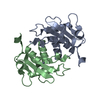
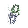
Evidence: gel filtration, This fragment of beta4 is a monomer (by SAXS, gel-filtration, and EPR). When BP230 binds to beta4, the latter remains monomeric (as judged by EPR)
Type
Name
Symmetry operation
Number
identity operation
1_555
x,y,z
1
Buried area
3640 Å2
ΔGint
-20 kcal/mol
Surface area
11250 Å2
Method
PISA
Unit cell
Length a, b, c (Å)
105.580, 59.520, 42.400
Angle α, β, γ (deg.)
90.00, 113.50, 90.00
Int Tables number
5
Space group name H-M
C121
Components on special symmetry positions
ID
Model
Components
1
1
A-1832-
HOH
-
Components
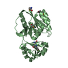
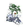
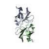
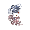
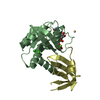
#1: Protein
Integrinbeta-4 / GP150
Mass: 23438.285 Da / Num. of mol.: 1 / Mutation: T1663R Source method: isolated from a genetically manipulated source Source: (gene. exp.) Homo sapiens (human) / Gene: ITGB4 / Plasmid: Modified pET15b / Production host: Escherichia coli BL21(DE3) (bacteria) / References: UniProt: P16144
In the structure databanks used in Yorodumi, some data are registered as the other names, "COVID-19 virus" and "2019-nCoV". Here are the details of the virus and the list of structure data.
Jan 31, 2019. EMDB accession codes are about to change! (news from PDBe EMDB page)
EMDB accession codes are about to change! (news from PDBe EMDB page)
The allocation of 4 digits for EMDB accession codes will soon come to an end. Whilst these codes will remain in use, new EMDB accession codes will include an additional digit and will expand incrementally as the available range of codes is exhausted. The current 4-digit format prefixed with “EMD-” (i.e. EMD-XXXX) will advance to a 5-digit format (i.e. EMD-XXXXX), and so on. It is currently estimated that the 4-digit codes will be depleted around Spring 2019, at which point the 5-digit format will come into force.
The EM Navigator/Yorodumi systems omit the EMD- prefix.
Related info.:Q: What is EMD? / ID/Accession-code notation in Yorodumi/EM Navigator
Yorodumi is a browser for structure data from EMDB, PDB, SASBDB, etc.
This page is also the successor to EM Navigator detail page, and also detail information page/front-end page for Omokage search.
The word "yorodu" (or yorozu) is an old Japanese word meaning "ten thousand". "mi" (miru) is to see.
Related info.:EMDB / PDB / SASBDB / Comparison of 3 databanks / Yorodumi Search / Aug 31, 2016. New EM Navigator & Yorodumi / Yorodumi Papers / Jmol/JSmol / Function and homology information / Changes in new EM Navigator and Yorodumi
 Movie
Movie Controller

 Yorodumi
 Open data
 Basic information
 Components
 Keywords
 Function and homology information
 Homo sapiens (human)
 X-RAY DIFFRACTION /
  Authors
 Spain, 2items
  Citation
 Structure visualization
 Downloads & links
 Other downloads


 PDBj
PDBj


 Assembly


 Mass: 92.094 Da / Num. of mol.: 2 / Source method: obtained synthetically / Formula: C3H8O3
 Mass: 18.015 Da / Num. of mol.: 105 / Source method: isolated from a natural source / Formula: H2O
 Sample preparation
 Processing