 Movie
Movie Controller

[English] 日本語
 Yorodumi
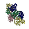
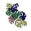
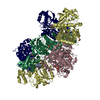
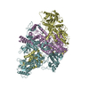
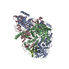
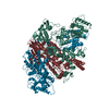
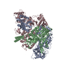
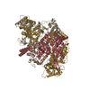
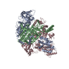
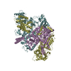
- PDB-6grg: E. coli Microcin synthetase McbBCD complex with pro-MccB17, ADP a... -
+ Open data

- Basic information
| Entry | Database: PDB / ID: 6grg | |||||||||||||||
|---|---|---|---|---|---|---|---|---|---|---|---|---|---|---|---|---|
| Title | E. coli Microcin synthetase McbBCD complex with pro-MccB17, ADP and phosphate bound | |||||||||||||||
 Components Components |
| |||||||||||||||
 Keywords Keywords | BIOSYNTHETIC PROTEIN / microcin / DNA gyrase / heterocyclization / posttranslational modification | |||||||||||||||
| Function / homology |  Function and homology information Function and homology informationnegative regulation of DNA replication / antibiotic biosynthetic process / killing of cells of another organism / oxidoreductase activity / defense response to bacterium / cytoplasm Similarity search - Function | |||||||||||||||
| Biological species |  | |||||||||||||||
| Method |  X-RAY DIFFRACTION / SYNCHROTRON / FOURIER SYNTHESIS / Resolution: 2.35 Å X-RAY DIFFRACTION / SYNCHROTRON / FOURIER SYNTHESIS / Resolution: 2.35 Å | |||||||||||||||
 Authors Authors | Ghilarov, D. / Stevenson, C.E.M. / Travin, D.Y. / Piskunova, J. / Serebryakova, M. / Maxwell, A. / Lawson, D.M. / Severinov, K. | |||||||||||||||
| Funding support |  Poland, Poland,  Russian Federation, Russian Federation,  United Kingdom, 4items United Kingdom, 4items
| |||||||||||||||
 Citation Citation | Journal: Mol. Cell / Year: 2019 Title: Architecture of Microcin B17 Synthetase: An Octameric Protein Complex Converting a Ribosomally Synthesized Peptide into a DNA Gyrase Poison. Authors: Ghilarov, D. / Stevenson, C.E.M. / Travin, D.Y. / Piskunova, J. / Serebryakova, M. / Maxwell, A. / Lawson, D.M. / Severinov, K. | |||||||||||||||
| History |
|
- Structure visualization
| Structure viewer | Molecule: MolmilJmol/JSmol |
|---|
- Downloads & links
-Download
| PDBx/mmCIF format | 6grg.cif.gz | 521.3 KB | Display | PDBx/mmCIF format |
|---|---|---|---|---|
| PDB format | pdb6grg.ent.gz | 420.7 KB | Display | PDB format |
| PDBx/mmJSON format | 6grg.json.gz | Tree view | PDBx/mmJSON format | |
| Others |  Other downloads Other downloads |
-Validation report
| Arichive directory | https://data.pdbj.org/pub/pdb/validation_reports/gr/6grgftp://data.pdbj.org/pub/pdb/validation_reports/gr/6grg | HTTPS FTP |
|---|
-Related structure data
| Related structure data |  6gosSC  6grhC  6griC S: Starting model for refinement C: citing same article ( |
|---|---|
| Similar structure data |
-Links
 PDBj
PDBj- Assembly
| Deposited unit | 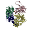
| ||||||||
|---|---|---|---|---|---|---|---|---|---|
| 1 | 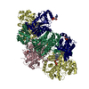
| ||||||||
| Unit cell |
|
-Components
-Protein , 1 types, 1 molecules A
| #1: Protein | Mass: 6853.401 Da / Num. of mol.: 1 Source method: isolated from a genetically manipulated source Details: The McbA protein was expressed with an eight-residue nickel affinity tag with sequence MGHHHHHH appended to the N-terminus of the full-length amino acid sequence. It was subsequently post- ...Details: The McbA protein was expressed with an eight-residue nickel affinity tag with sequence MGHHHHHH appended to the N-terminus of the full-length amino acid sequence. It was subsequently post-translationally modified by the McbBCD complex to yield nine heterocycles, where F6N equals oxazole and is derived from Gly-Ser, F75 equals thiazole and is derived from Gly-Cys, OTZ equals oxazole-thiazole and is derived from Gly-Ser-Cys, TOZ equals thiazole-oxazole and is derived from Gly-Cys-Ser. Each mono- or bis-heterocycle (where present) was treated as a pseudo-amino acid for refinement purposes. The numbering system used in this PDB file relates to the processed peptide where each pseudo-amino acid is treated as a single residue making the overall sequence nine residues shorter. Source: (gene. exp.) Gene: mcbA / Plasmid: pBAD-McbABCDEFG / Production host: |
|---|
-Microcin B17-processing protein ... , 3 types, 4 molecules 12CD
| #2: Protein | Mass: 34032.363 Da / Num. of mol.: 2 Source method: isolated from a genetically manipulated source Source: (gene. exp.) Gene: mcbB / Plasmid: pBAD-McbABCDEFG / Production host: #3: Protein | | Mass: 30789.057 Da / Num. of mol.: 1 Source method: isolated from a genetically manipulated source Source: (gene. exp.) Gene: mcbC / Plasmid: pBAD-McbABCDEFG / Production host: #4: Protein | | Mass: 44963.973 Da / Num. of mol.: 1 Source method: isolated from a genetically manipulated source Details: There is a T171R substitution relative to UniProtKB sequence Source: (gene. exp.) Gene: mcbD / Plasmid: pBAD-McbABCDEFG / Production host: |
|---|
-Non-polymers , 11 types, 240 molecules 


| #5: Chemical | ChemComp-FMN /  Mass: 456.344 Da / Num. of mol.: 1 / Source method: obtained synthetically / Formula: C17H21N4O9P Mass: 456.344 Da / Num. of mol.: 1 / Source method: obtained synthetically / Formula: C17H21N4O9P | ||||||||||||||||||
|---|---|---|---|---|---|---|---|---|---|---|---|---|---|---|---|---|---|---|---|
| #6: Chemical |  Mass: 65.409 Da / Num. of mol.: 2 / Source method: obtained synthetically / Formula: Zn Mass: 65.409 Da / Num. of mol.: 2 / Source method: obtained synthetically / Formula: Zn#7: Chemical | ChemComp-SO4 / |  Mass: 96.063 Da / Num. of mol.: 1 / Source method: obtained synthetically / Formula: SO4 Mass: 96.063 Da / Num. of mol.: 1 / Source method: obtained synthetically / Formula: SO4#8: Chemical | ChemComp-GOL / |  Mass: 92.094 Da / Num. of mol.: 1 / Source method: obtained synthetically / Formula: C3H8O3 Mass: 92.094 Da / Num. of mol.: 1 / Source method: obtained synthetically / Formula: C3H8O3#9: Chemical | ChemComp-ATP / |  Mass: 507.181 Da / Num. of mol.: 1 / Source method: obtained synthetically / Formula: C10H16N5O13P3 / Comment: ATP, energy-carrying molecule*YM Mass: 507.181 Da / Num. of mol.: 1 / Source method: obtained synthetically / Formula: C10H16N5O13P3 / Comment: ATP, energy-carrying molecule*YM#10: Chemical |  Mass: 62.068 Da / Num. of mol.: 2 / Source method: obtained synthetically / Formula: C2H6O2 Mass: 62.068 Da / Num. of mol.: 2 / Source method: obtained synthetically / Formula: C2H6O2#11: Chemical | ChemComp-ADP / |  Mass: 427.201 Da / Num. of mol.: 1 / Source method: obtained synthetically / Formula: C10H15N5O10P2 / Comment: ADP, energy-carrying molecule*YM Mass: 427.201 Da / Num. of mol.: 1 / Source method: obtained synthetically / Formula: C10H15N5O10P2 / Comment: ADP, energy-carrying molecule*YM#12: Chemical | ChemComp-PO4 / |  Mass: 94.971 Da / Num. of mol.: 1 / Source method: obtained synthetically / Formula: PO4 Mass: 94.971 Da / Num. of mol.: 1 / Source method: obtained synthetically / Formula: PO4#13: Chemical |  Mass: 24.305 Da / Num. of mol.: 3 / Source method: obtained synthetically / Formula: Mg Mass: 24.305 Da / Num. of mol.: 3 / Source method: obtained synthetically / Formula: Mg#14: Chemical | ChemComp-CL / |  Mass: 35.453 Da / Num. of mol.: 1 / Source method: obtained synthetically / Formula: Cl Mass: 35.453 Da / Num. of mol.: 1 / Source method: obtained synthetically / Formula: Cl#15: Water | ChemComp-HOH / | Mass: 18.015 Da / Num. of mol.: 226 / Source method: isolated from a natural source / Formula: H2O |
-Details
| Has protein modification | Y |
|---|
-Experimental details
-Experiment
| Experiment | Method: X-RAY DIFFRACTION / Number of used crystals: 1 |
|---|
- Sample preparation
| Crystal | Density Matthews: 2.17 Å3/Da / Density % sol: 43.21 % |
|---|---|
| Crystal grow | Temperature: 293 K / Method: vapor diffusion, sitting drop / Details: NULL |
-Data collection
| Diffraction | Mean temperature: 100 K | ||||||||||||||||||||||||
|---|---|---|---|---|---|---|---|---|---|---|---|---|---|---|---|---|---|---|---|---|---|---|---|---|---|
| Diffraction source | Source: SYNCHROTRON / Site: Diamond / Beamline: I04 / Wavelength: 0.9795 Å | ||||||||||||||||||||||||
| Detector | Type: DECTRIS PILATUS3 6M / Detector: PIXEL / Date: Feb 7, 2016 | ||||||||||||||||||||||||
| Radiation | Protocol: SINGLE WAVELENGTH / Monochromatic (M) / Laue (L): M / Scattering type: x-ray | ||||||||||||||||||||||||
| Radiation wavelength | Wavelength: 0.9795 Å / Relative weight: 1 | ||||||||||||||||||||||||
| Reflection | Resolution: 2.35→86.76 Å / Num. obs: 53732 / % possible obs: 99.9 % / Redundancy: 7.6 % / CC1/2: 0.998 / Rmerge(I) obs: 0.14 / Rpim(I) all: 0.054 / Rrim(I) all: 0.15 / Net I/σ(I): 11.6 / Num. measured all: 409359 / Scaling rejects: 2 | ||||||||||||||||||||||||
| Reflection shell | Diffraction-ID: 1
|
- Processing
| Software |
| ||||||||||||||||||||||||||||||||||||||||||||||||||||||||||||||||||||||||||||||||||||||||||||||||||||||||||||||||||||||||||||||||||||||||||||||||||||||
|---|---|---|---|---|---|---|---|---|---|---|---|---|---|---|---|---|---|---|---|---|---|---|---|---|---|---|---|---|---|---|---|---|---|---|---|---|---|---|---|---|---|---|---|---|---|---|---|---|---|---|---|---|---|---|---|---|---|---|---|---|---|---|---|---|---|---|---|---|---|---|---|---|---|---|---|---|---|---|---|---|---|---|---|---|---|---|---|---|---|---|---|---|---|---|---|---|---|---|---|---|---|---|---|---|---|---|---|---|---|---|---|---|---|---|---|---|---|---|---|---|---|---|---|---|---|---|---|---|---|---|---|---|---|---|---|---|---|---|---|---|---|---|---|---|---|---|---|---|---|---|---|
| Refinement | Method to determine structure: FOURIER SYNTHESIS Starting model: 6GOS Resolution: 2.35→86.76 Å / Cor.coef. Fo:Fc: 0.97 / Cor.coef. Fo:Fc free: 0.941 / SU B: 20.463 / SU ML: 0.225 / SU R Cruickshank DPI: 0.4202 / Cross valid method: THROUGHOUT / σ(F): 0 / ESU R: 0.407 / ESU R Free: 0.243 Details: HYDROGENS HAVE BEEN ADDED IN THE RIDING POSITIONS U VALUES : WITH TLS ADDED
| ||||||||||||||||||||||||||||||||||||||||||||||||||||||||||||||||||||||||||||||||||||||||||||||||||||||||||||||||||||||||||||||||||||||||||||||||||||||
| Solvent computation | Ion probe radii: 0.7 Å / Shrinkage radii: 0.7 Å / VDW probe radii: 1 Å | ||||||||||||||||||||||||||||||||||||||||||||||||||||||||||||||||||||||||||||||||||||||||||||||||||||||||||||||||||||||||||||||||||||||||||||||||||||||
| Displacement parameters | Biso max: 144.28 Å2 / Biso mean: 53.476 Å2 / Biso min: 27.57 Å2
| ||||||||||||||||||||||||||||||||||||||||||||||||||||||||||||||||||||||||||||||||||||||||||||||||||||||||||||||||||||||||||||||||||||||||||||||||||||||
| Refinement step | Cycle: final / Resolution: 2.35→86.76 Å
| ||||||||||||||||||||||||||||||||||||||||||||||||||||||||||||||||||||||||||||||||||||||||||||||||||||||||||||||||||||||||||||||||||||||||||||||||||||||
| Refine LS restraints |
| ||||||||||||||||||||||||||||||||||||||||||||||||||||||||||||||||||||||||||||||||||||||||||||||||||||||||||||||||||||||||||||||||||||||||||||||||||||||
| LS refinement shell | Resolution: 2.35→2.411 Å / Rfactor Rfree error: 0 / Total num. of bins used: 20
| ||||||||||||||||||||||||||||||||||||||||||||||||||||||||||||||||||||||||||||||||||||||||||||||||||||||||||||||||||||||||||||||||||||||||||||||||||||||
| Refinement TLS params. | Method: refined / Refine-ID: X-RAY DIFFRACTION
| ||||||||||||||||||||||||||||||||||||||||||||||||||||||||||||||||||||||||||||||||||||||||||||||||||||||||||||||||||||||||||||||||||||||||||||||||||||||
| Refinement TLS group |
|
