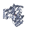
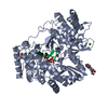
Entry Database : PDB / ID : 6gp9Title Structural studies of hepatitis C virus non-structural protein-5b of genotype 4a RNA-directed RNA polymerase Keywords / / / Function / homology Function Domain/homology Component
/ / / / / / / / / / / / / / / Biological species Method / / / Resolution : 3.1 Å Authors Janowski, R. / Geber, H. / Protzer, U. / Niessing, D. Journal : Biorxiv / Year : 2022Title : Structural studies of hepatitis C virus non-structural protein-5b of genotype 4aAuthors : Gaber, H. / Niessing, D. / Protzer, U. / Janowski, R. History Deposition Jun 5, 2018 Deposition site / Processing site Revision 1.0 Sep 30, 2020 Provider / Type Revision 1.1 Apr 13, 2022 Group / Category / citation_author / database_2Item _citation.country / _citation.journal_abbrev ... _citation.country / _citation.journal_abbrev / _citation.journal_id_CSD / _citation.pdbx_database_id_DOI / _citation.year / _citation_author.name / _database_2.pdbx_DOI / _database_2.pdbx_database_accession Revision 1.2 Jan 17, 2024 Group / Database references / Refinement descriptionCategory chem_comp_atom / chem_comp_bond ... chem_comp_atom / chem_comp_bond / citation / pdbx_initial_refinement_model Item Revision 1.3 Oct 9, 2024 Group / Category / pdbx_modification_feature
Show all Show less
 Movie
Movie Controller

 Yorodumi
 Open data
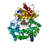
 Basic information
 Components
 Keywords
 Function and homology information
 Hepacivirus C
 X-RAY DIFFRACTION /
  Authors
 Citation
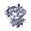
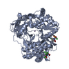
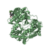
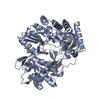
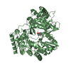
 Structure visualization
 Downloads & links
 Other downloads
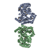

 PDBj
PDBj
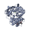
 Assembly
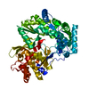

 Mass: 18.015 Da / Num. of mol.: 16 / Source method: isolated from a natural source / Formula: H2O
 Sample preparation
 / Beamline: MASSIF-3 / Wavelength: 0.965 Å
 Processing