 ムービー
ムービー コントローラー


+ データを開く

- 基本情報
基本情報
| 登録情報 | データベース: PDB / ID: 6gi6 | ||||||
|---|---|---|---|---|---|---|---|
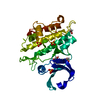
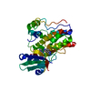
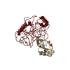
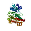
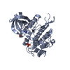
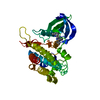
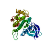
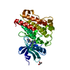
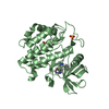
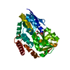
| タイトル | Crystal structure of the ACVR1 (ALK2) kinase in complex with a Quinazolinone based ALK2 inhibitor with a 5-methyl core. | ||||||
 要素 要素 | Activin receptor type-1 | ||||||
 キーワード キーワード | SIGNALING PROTEIN / Kinase / BMP / inhibitor / signalling | ||||||
| 機能・相同性 |  機能・相同性情報 機能・相同性情報endocardial cushion cell fate commitment / positive regulation of determination of dorsal identity / atrial septum primum morphogenesis / acute inflammatory response / mitral valve morphogenesis / BMP receptor complex / BMP receptor activity / endocardial cushion fusion / cardiac muscle cell fate commitment / positive regulation of cardiac epithelial to mesenchymal transition ...endocardial cushion cell fate commitment / positive regulation of determination of dorsal identity / atrial septum primum morphogenesis / acute inflammatory response / mitral valve morphogenesis / BMP receptor complex / BMP receptor activity / endocardial cushion fusion / cardiac muscle cell fate commitment / positive regulation of cardiac epithelial to mesenchymal transition / transforming growth factor beta receptor activity, type I / activin receptor activity, type I / activin receptor complex / endocardial cushion formation / receptor protein serine/threonine kinase / cellular response to BMP stimulus / activin binding / embryonic heart tube morphogenesis / transmembrane receptor protein serine/threonine kinase activity / activin receptor signaling pathway / dorsal/ventral pattern formation / negative regulation of activin receptor signaling pathway / transforming growth factor beta binding / atrioventricular valve morphogenesis / ventricular septum morphogenesis / negative regulation of G1/S transition of mitotic cell cycle / positive regulation of bone mineralization / SMAD binding / positive regulation of osteoblast differentiation / peptide hormone binding / positive regulation of intracellular signal transduction / positive regulation of SMAD protein signal transduction / regulation of ossification / BMP signaling pathway / negative regulation of signal transduction / transforming growth factor beta receptor signaling pathway / protein tyrosine kinase binding / negative regulation of extrinsic apoptotic signaling pathway / cellular response to growth factor stimulus / osteoblast differentiation / apical part of cell / heart development / cell differentiation / protein kinase activity / positive regulation of cell migration / cadherin binding / protein serine/threonine kinase activity / positive regulation of DNA-templated transcription / positive regulation of transcription by RNA polymerase II / protein homodimerization activity / ATP binding / metal ion binding / plasma membrane 類似検索 - 分子機能 | ||||||
| 生物種 |  Homo sapiens (ヒト) Homo sapiens (ヒト) | ||||||
| 手法 |  X線回折 / シンクロトロン / 分子置換 / 解像度: 1.98 Å X線回折 / シンクロトロン / 分子置換 / 解像度: 1.98 Å | ||||||
 データ登録者 データ登録者 | Williams, E. / Hudson, L. / Bezerra, G.A. / Sorrell, F. / Mahajan, P. / Kupinska, K. / Hoelder, S. / Burgess-Brown, N. / von Delft, F. / Arrowsmith, C.H. ...Williams, E. / Hudson, L. / Bezerra, G.A. / Sorrell, F. / Mahajan, P. / Kupinska, K. / Hoelder, S. / Burgess-Brown, N. / von Delft, F. / Arrowsmith, C.H. / Edwards, A.M. / Bountra, C. / Bullock, A.N. | ||||||
 引用 引用 | ジャーナル: J. Med. Chem. / 年: 2018 タイトル: Novel Quinazolinone Inhibitors of ALK2 Flip between Alternate Binding Modes: Structure-Activity Relationship, Structural Characterization, Kinase Profiling, and Cellular Proof of Concept. 著者: Hudson, L. / Mui, J. / Vazquez, S. / Carvalho, D.M. / Williams, E. / Jones, C. / Bullock, A.N. / Hoelder, S. | ||||||
| 履歴 |
|
- 構造の表示
構造の表示
| 構造ビューア | 分子: MolmilJmol/JSmol |
|---|
- ダウンロードとリンク
-ダウンロード
| PDBx/mmCIF形式 | 6gi6.cif.gz | 139.1 KB | 表示 | PDBx/mmCIF形式 |
|---|---|---|---|---|
| PDB形式 | pdb6gi6.ent.gz | 106.5 KB | 表示 | PDB形式 |
| PDBx/mmJSON形式 | 6gi6.json.gz | ツリー表示 | PDBx/mmJSON形式 | |
| その他 |  その他のダウンロード その他のダウンロード |
-検証レポート
| アーカイブディレクトリ | https://data.pdbj.org/pub/pdb/validation_reports/gi/6gi6ftp://data.pdbj.org/pub/pdb/validation_reports/gi/6gi6 | HTTPS FTP |
|---|
-関連構造データ


-リンク
 PDBj
PDBj
- 集合体
集合体
| 登録構造単位 | 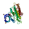
| ||||||||||||
|---|---|---|---|---|---|---|---|---|---|---|---|---|---|
| 1 |
| ||||||||||||
| 単位格子 |
|
-要素
| #1: タンパク質 | 分子量: 34537.633 Da / 分子数: 1 / 変異: Q207D / 由来タイプ: 組換発現 / 由来: (組換発現) Homo sapiens (ヒト) / 遺伝子: ACVR1, ACVRLK2発現宿主:   Spodoptera frugiperda (ツマジロクサヨトウ) Spodoptera frugiperda (ツマジロクサヨトウ)株 (発現宿主): Sf9 参照: UniProt: Q04771, receptor protein serine/threonine kinase | ||||
|---|---|---|---|---|---|
| #2: 化合物 | ChemComp-EZB /   分子量: 287.315 Da / 分子数: 1 / 由来タイプ: 合成 / 式: C18H13N3O 分子量: 287.315 Da / 分子数: 1 / 由来タイプ: 合成 / 式: C18H13N3O | ||||
| #3: 化合物 | ChemComp-SO4 /   分子量: 96.063 Da / 分子数: 4 / 由来タイプ: 合成 / 式: SO4 分子量: 96.063 Da / 分子数: 4 / 由来タイプ: 合成 / 式: SO4#4: 化合物 | ChemComp-EDO /   分子量: 62.068 Da / 分子数: 10 / 由来タイプ: 合成 / 式: C2H6O2 分子量: 62.068 Da / 分子数: 10 / 由来タイプ: 合成 / 式: C2H6O2#5: 水 | ChemComp-HOH / |  分子量: 18.015 Da / 分子数: 149 / 由来タイプ: 天然 / 式: H2O 分子量: 18.015 Da / 分子数: 149 / 由来タイプ: 天然 / 式: H2O |
-実験情報
-実験
| 実験 | 手法: X線回折 / 使用した結晶の数: 1 |
|---|
- 試料調製
試料調製
| 結晶 | マシュー密度: 2.49 Å3/Da / 溶媒含有率: 50.67 % |
|---|---|
| 結晶化 | 温度: 277 K / 手法: 蒸気拡散法, シッティングドロップ法 / pH: 6.5 詳細: 1.5M ammonium sulfate, 0.1M sodium chloride, 0.1M bis-tris pH 6.5 |
-データ収集
| 回折 | 平均測定温度: 100 K |
|---|---|
| 放射光源 | 由来: シンクロトロン / サイト: Diamond  / ビームライン: I02 / 波長: 0.9795 Å / ビームライン: I02 / 波長: 0.9795 Å |
| 検出器 | タイプ: DECTRIS PILATUS 6M / 検出器: PIXEL / 日付: 2016年1月5日 詳細: Kirkpatrick Baez (KB) bimorph mirror pair for horizontal and vertical focussing |
| 放射 | モノクロメーター: Double Crystal / プロトコル: SINGLE WAVELENGTH / 単色(M)・ラウエ(L): M / 散乱光タイプ: x-ray |
| 放射波長 | 波長: 0.9795 Å / 相対比: 1 |
| 反射 | 解像度: 1.98→73 Å / Num. obs: 26321 / % possible obs: 99.7 % / Observed criterion σ(I): 1.5 / 冗長度: 4.2 % / Biso Wilson estimate: 8.1 Å2 / CC1/2: 0.943 / Rmerge(I) obs: 0.283 / Rpim(I) all: 0.152 / Rrim(I) all: 0.323 / Net I/σ(I): 4.1 |
| 反射 シェル | 解像度: 1.98→2.03 Å / 冗長度: 4.4 % / Rmerge(I) obs: 0.899 / Mean I/σ(I) obs: 1.6 / Num. unique obs: 1825 / CC1/2: 0.239 / Rpim(I) all: 0.477 / Rrim(I) all: 1.023 / % possible all: 99.7 |
- 解析
解析
| ソフトウェア |
| ||||||||||||||||||||
|---|---|---|---|---|---|---|---|---|---|---|---|---|---|---|---|---|---|---|---|---|---|
| 精密化 | 構造決定の手法: 分子置換 開始モデル: 3H9R 解像度: 1.98→57.28 Å / 交差検証法: FREE R-VALUE
| ||||||||||||||||||||
| 精密化ステップ | サイクル: LAST / 解像度: 1.98→57.28 Å
|
