 Movie
Movie Controller

[English] 日本語
 Yorodumi
- PDB-6ghv: Structure of a DC-SIGN CRD in complex with high affinity glycomimetic. -
+ Open data

- Basic information
| Entry | Database: PDB / ID: 6ghv | ||||||
|---|---|---|---|---|---|---|---|
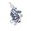
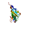
| Title | Structure of a DC-SIGN CRD in complex with high affinity glycomimetic. | ||||||
 Components Components | CD209 antigen | ||||||
 Keywords Keywords | SUGAR BINDING PROTEIN / DC-SIGN / Inhibitor | ||||||
| Function / homology |  Function and homology information Function and homology informationB cell adhesion / immature T cell proliferation / cell-cell recognition / intracellular transport of virus / cell adhesion receptor activity / peptide antigen transport / dendritic cell migration / Butyrophilin (BTN) family interactions / positive regulation of viral life cycle / virion binding ...B cell adhesion / immature T cell proliferation / cell-cell recognition / intracellular transport of virus / cell adhesion receptor activity / peptide antigen transport / dendritic cell migration / Butyrophilin (BTN) family interactions / positive regulation of viral life cycle / virion binding / heterophilic cell-cell adhesion / leukocyte cell-cell adhesion / antigen processing and presentation / pattern recognition receptor activity / RSV-host interactions / D-mannose binding / regulation of T cell proliferation / positive regulation of T cell proliferation / CD209 (DC-SIGN) signaling / viral genome replication / peptide antigen binding / endocytosis / host cell / carbohydrate binding / virus receptor activity / adaptive immune response / intracellular signal transduction / immune response / external side of plasma membrane / innate immune response / symbiont entry into host cell / virion attachment to host cell / cell surface / extracellular region / membrane / metal ion binding / plasma membrane / cytoplasm Similarity search - Function | ||||||
| Biological species |  Homo sapiens (human) Homo sapiens (human) | ||||||
| Method |  X-RAY DIFFRACTION / SYNCHROTRON / MOLECULAR REPLACEMENT / molecular replacement / Resolution: 2.1 Å X-RAY DIFFRACTION / SYNCHROTRON / MOLECULAR REPLACEMENT / molecular replacement / Resolution: 2.1 Å | ||||||
 Authors Authors | Thepaut, M. / Achilli, S. / Medve, L. / Bernardi, A. / Fieschi, F. | ||||||
| Funding support |  France, 1items France, 1items
| ||||||
 Citation Citation | Journal: Chemistry / Year: 2019 Title: Enhancing Potency and Selectivity of a DC-SIGN Glycomimetic Ligand by Fragment-Based Design: Structural Basis. Authors: Medve, L. / Achilli, S. / Guzman-Caldentey, J. / Thepaut, M. / Senaldi, L. / Le Roy, A. / Sattin, S. / Ebel, C. / Vives, C. / Martin-Santamaria, S. / Bernardi, A. / Fieschi, F. | ||||||
| History |
|
- Structure visualization
| Structure viewer | Molecule: MolmilJmol/JSmol |
|---|
- Downloads & links
-Download
| PDBx/mmCIF format | 6ghv.cif.gz | 211.3 KB | Display | PDBx/mmCIF format |
|---|---|---|---|---|
| PDB format | pdb6ghv.ent.gz | 164.6 KB | Display | PDB format |
| PDBx/mmJSON format | 6ghv.json.gz | Tree view | PDBx/mmJSON format | |
| Others |  Other downloads Other downloads |
-Validation report
| Arichive directory | https://data.pdbj.org/pub/pdb/validation_reports/gh/6ghvftp://data.pdbj.org/pub/pdb/validation_reports/gh/6ghv | HTTPS FTP |
|---|
-Related structure data
| Related structure data | 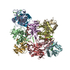 1k9iS S: Starting model for refinement |
|---|---|
| Similar structure data |


-Links
 PDBj
PDBj


- Assembly
| Deposited unit | 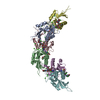
| ||||||||
|---|---|---|---|---|---|---|---|---|---|
| 1 | 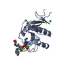
| ||||||||
| 2 | 
| ||||||||
| 3 | 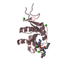
| ||||||||
| 4 | 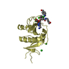
| ||||||||
| 5 | 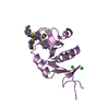
| ||||||||
| 6 | 
| ||||||||
| Unit cell |
|
-Components
| #1: Protein | Mass: 17746.582 Da / Num. of mol.: 6 Source method: isolated from a genetically manipulated source Source: (gene. exp.) Homo sapiens (human) / Gene: CD209, CLEC4L / Plasmid: pET30 / Production host:  #2: Chemical | ChemComp-EZ8 / [   Mass: 748.243 Da / Num. of mol.: 6 / Source method: obtained synthetically / Formula: C35H48ClN6O10 / Feature type: SUBJECT OF INVESTIGATION Mass: 748.243 Da / Num. of mol.: 6 / Source method: obtained synthetically / Formula: C35H48ClN6O10 / Feature type: SUBJECT OF INVESTIGATION#3: Chemical | ChemComp-CA /   Mass: 40.078 Da / Num. of mol.: 18 / Source method: isolated from a natural source / Formula: Ca Mass: 40.078 Da / Num. of mol.: 18 / Source method: isolated from a natural source / Formula: Ca#4: Chemical | ChemComp-CL /   Mass: 35.453 Da / Num. of mol.: 14 / Source method: obtained synthetically / Formula: Cl Mass: 35.453 Da / Num. of mol.: 14 / Source method: obtained synthetically / Formula: Cl#5: Water | ChemComp-HOH / |  Mass: 18.015 Da / Num. of mol.: 660 / Source method: isolated from a natural source / Formula: H2O Mass: 18.015 Da / Num. of mol.: 660 / Source method: isolated from a natural source / Formula: H2OHas protein modification | Y | |
|---|
-Experimental details
-Experiment
| Experiment | Method: X-RAY DIFFRACTION / Number of used crystals: 1 |
|---|
- Sample preparation
| Crystal | Density Matthews: 3.15 Å3/Da / Density % sol: 60.9 % |
|---|---|
| Crystal grow | Temperature: 293 K / Method: vapor diffusion, hanging drop Details: 20% PEG 3350, 200mM Mg(NO3)2, 100mM MES pH6. Protein sample: 150mM NaCl, 4mM CaCl2, 25mM Tris pH8, 2% DMSO, 3.25 mM ligand and 5.54mg/mL protein. |
-Data collection
| Diffraction | Mean temperature: 100 K | ||||||||||||||||||||||||||||||||||||||||||||||||||||||||||||||||||||||||||||||||||||||||||||||||||||||||||||||||||||||||
|---|---|---|---|---|---|---|---|---|---|---|---|---|---|---|---|---|---|---|---|---|---|---|---|---|---|---|---|---|---|---|---|---|---|---|---|---|---|---|---|---|---|---|---|---|---|---|---|---|---|---|---|---|---|---|---|---|---|---|---|---|---|---|---|---|---|---|---|---|---|---|---|---|---|---|---|---|---|---|---|---|---|---|---|---|---|---|---|---|---|---|---|---|---|---|---|---|---|---|---|---|---|---|---|---|---|---|---|---|---|---|---|---|---|---|---|---|---|---|---|---|---|
| Diffraction source | Source: SYNCHROTRON / Site: ESRF / Beamline: MASSIF-1 / Wavelength: 0.966 Å | ||||||||||||||||||||||||||||||||||||||||||||||||||||||||||||||||||||||||||||||||||||||||||||||||||||||||||||||||||||||||
| Detector | Type: DECTRIS PILATUS3 2M / Detector: PIXEL / Date: Feb 27, 2018 | ||||||||||||||||||||||||||||||||||||||||||||||||||||||||||||||||||||||||||||||||||||||||||||||||||||||||||||||||||||||||
| Radiation | Protocol: SINGLE WAVELENGTH / Monochromatic (M) / Laue (L): M / Scattering type: x-ray | ||||||||||||||||||||||||||||||||||||||||||||||||||||||||||||||||||||||||||||||||||||||||||||||||||||||||||||||||||||||||
| Radiation wavelength | Wavelength: 0.966 Å / Relative weight: 1 | ||||||||||||||||||||||||||||||||||||||||||||||||||||||||||||||||||||||||||||||||||||||||||||||||||||||||||||||||||||||||
| Reflection | Resolution: 2.1→40 Å / Num. obs: 65455 / % possible obs: 98.4 % / Redundancy: 3.087 % / Biso Wilson estimate: 34.771 Å2 / CC1/2: 0.993 / Rmerge(I) obs: 0.099 / Rrim(I) all: 0.12 / Χ2: 1.005 / Net I/σ(I): 8.47 | ||||||||||||||||||||||||||||||||||||||||||||||||||||||||||||||||||||||||||||||||||||||||||||||||||||||||||||||||||||||||
| Reflection shell | Diffraction-ID: 1
|
-Phasing
| Phasing | Method: molecular replacement | ||||||
|---|---|---|---|---|---|---|---|
| Phasing MR | R rigid body: 0.583
|
- Processing
| Software |
| ||||||||||||||||||||||||||||||||||||||||||||||||||||||||||||||||||||||||||||||||||||||||||||||||||||||||||||||||||||||||||||||||||||||||||||||||||||||||||||||||||||||||||||||||||||||
|---|---|---|---|---|---|---|---|---|---|---|---|---|---|---|---|---|---|---|---|---|---|---|---|---|---|---|---|---|---|---|---|---|---|---|---|---|---|---|---|---|---|---|---|---|---|---|---|---|---|---|---|---|---|---|---|---|---|---|---|---|---|---|---|---|---|---|---|---|---|---|---|---|---|---|---|---|---|---|---|---|---|---|---|---|---|---|---|---|---|---|---|---|---|---|---|---|---|---|---|---|---|---|---|---|---|---|---|---|---|---|---|---|---|---|---|---|---|---|---|---|---|---|---|---|---|---|---|---|---|---|---|---|---|---|---|---|---|---|---|---|---|---|---|---|---|---|---|---|---|---|---|---|---|---|---|---|---|---|---|---|---|---|---|---|---|---|---|---|---|---|---|---|---|---|---|---|---|---|---|---|---|---|---|
| Refinement | Method to determine structure: MOLECULAR REPLACEMENT Starting model: 1K9I Resolution: 2.1→40 Å / Cor.coef. Fo:Fc: 0.96 / Cor.coef. Fo:Fc free: 0.925 / SU B: 5.856 / SU ML: 0.145 / Cross valid method: THROUGHOUT / ESU R: 0.184 / ESU R Free: 0.175 / Details: HYDROGENS HAVE BEEN ADDED IN THE RIDING POSITIONS
| ||||||||||||||||||||||||||||||||||||||||||||||||||||||||||||||||||||||||||||||||||||||||||||||||||||||||||||||||||||||||||||||||||||||||||||||||||||||||||||||||||||||||||||||||||||||
| Solvent computation | Ion probe radii: 0.8 Å / Shrinkage radii: 0.8 Å / VDW probe radii: 1.2 Å | ||||||||||||||||||||||||||||||||||||||||||||||||||||||||||||||||||||||||||||||||||||||||||||||||||||||||||||||||||||||||||||||||||||||||||||||||||||||||||||||||||||||||||||||||||||||
| Displacement parameters | Biso mean: 33.681 Å2
| ||||||||||||||||||||||||||||||||||||||||||||||||||||||||||||||||||||||||||||||||||||||||||||||||||||||||||||||||||||||||||||||||||||||||||||||||||||||||||||||||||||||||||||||||||||||
| Refinement step | Cycle: 1 / Resolution: 2.1→40 Å
| ||||||||||||||||||||||||||||||||||||||||||||||||||||||||||||||||||||||||||||||||||||||||||||||||||||||||||||||||||||||||||||||||||||||||||||||||||||||||||||||||||||||||||||||||||||||
| Refine LS restraints |
|