 Movie
Movie Controller


+ Open data

- Basic information
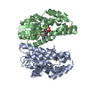
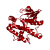
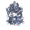
| Entry | Database: PDB / ID: 6ghf | ||||||
|---|---|---|---|---|---|---|---|
| Title | Crystal structure of a GST variant | ||||||
 Components Components | PvGmGSTUG | ||||||
 Keywords Keywords | BIOSYNTHETIC PROTEIN / gene shuffling / transferase | ||||||
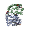
| Function / homology | Glutathione S-transferase Yfyf (Class Pi); Chain A, domain 2 - #10 / Glutathione S-transferase Yfyf (Class Pi); Chain A, domain 2 / Glutaredoxin / Glutaredoxin / Up-down Bundle / 3-Layer(aba) Sandwich / Mainly Alpha / Alpha Beta Function and homology information Function and homology information | ||||||
| Biological species | synthetic construct (others) | ||||||
| Method |  X-RAY DIFFRACTION / SYNCHROTRON / MOLECULAR REPLACEMENT / Resolution: 3.52 Å X-RAY DIFFRACTION / SYNCHROTRON / MOLECULAR REPLACEMENT / Resolution: 3.52 Å | ||||||
 Authors Authors | Papageorgiou, A.C. / Chronopoulou, E.G. / Labrou, N.E. | ||||||
 Citation Citation | Journal: Front Plant Sci / Year: 2018 Title: Expanding the Plant GSTome Through Directed Evolution: DNA Shuffling for the Generation of New Synthetic Enzymes With Engineered Catalytic and Binding Properties. Authors: Chronopoulou, E.G. / Papageorgiou, A.C. / Ataya, F. / Nianiou-Obeidat, I. / Madesis, P. / Labrou, N.E. | ||||||
| History |
|
- Structure visualization
| Structure viewer | Molecule: MolmilJmol/JSmol |
|---|
- Downloads & links
-Download
| PDBx/mmCIF format | 6ghf.cif.gz | 97.7 KB | Display | PDBx/mmCIF format |
|---|---|---|---|---|
| PDB format | pdb6ghf.ent.gz | 75.6 KB | Display | PDB format |
| PDBx/mmJSON format | 6ghf.json.gz | Tree view | PDBx/mmJSON format | |
| Others |  Other downloads Other downloads |
-Validation report
| Arichive directory | https://data.pdbj.org/pub/pdb/validation_reports/gh/6ghfftp://data.pdbj.org/pub/pdb/validation_reports/gh/6ghf | HTTPS FTP |
|---|
-Related structure data
| Related structure data |  4j2fS S: Starting model for refinement |
|---|---|
| Similar structure data |


-Links
 PDBj
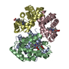
PDBj- Assembly
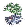
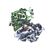
| Deposited unit | 
| ||||||||||||||||||
|---|---|---|---|---|---|---|---|---|---|---|---|---|---|---|---|---|---|---|---|
| 1 |
| ||||||||||||||||||
| Unit cell |
| ||||||||||||||||||
| Noncrystallographic symmetry (NCS) | NCS domain:
NCS domain segments: Component-ID: _ / Ens-ID: 1 / Beg auth comp-ID: GLU / Beg label comp-ID: GLU / End auth comp-ID: TYR / End label comp-ID: TYR / Refine code: _ / Auth seq-ID: 6 - 212 / Label seq-ID: 6 - 212
|
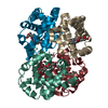
-Components
| #1: Protein | Mass: 26124.057 Da / Num. of mol.: 2 Source method: isolated from a genetically manipulated source Details: The gene expressing PvGmGSTUG was constructed by gene shuffling of a Phaseolus vulgaris GST and a Glycine max GST. Source: (gene. exp.) synthetic construct (others) / Gene: Synthetic / Production host:  |
|---|
-Experimental details
-Experiment
| Experiment | Method: X-RAY DIFFRACTION / Number of used crystals: 1 |
|---|
- Sample preparation
| Crystal | Density Matthews: 2.83 Å3/Da / Density % sol: 56.56 % / Description: Rods |
|---|---|
| Crystal grow | Temperature: 289 K / Method: vapor diffusion, hanging drop / pH: 7 Details: PEG 4000 20% (w/v), Na succinate 0.2 M, Hepes-NaOH 0.1 M |
-Data collection
| Diffraction | Mean temperature: 100 K |
|---|---|
| Diffraction source | Source: SYNCHROTRON / Site: ESRF  / Beamline: ID23-1 / Wavelength: 0.973 Å / Beamline: ID23-1 / Wavelength: 0.973 Å |
| Detector | Type: DECTRIS PILATUS3 6M / Detector: PIXEL / Date: Jun 24, 2014 |
| Radiation | Protocol: SINGLE WAVELENGTH / Monochromatic (M) / Laue (L): M / Scattering type: x-ray |
| Radiation wavelength | Wavelength: 0.973 Å / Relative weight: 1 |
| Reflection | Resolution: 3.5→56.87 Å / Num. obs: 7132 / % possible obs: 98.6 % / Redundancy: 4.8 % / CC1/2: 0.998 / Rrim(I) all: 0.099 / Net I/σ(I): 8.8 |
| Reflection shell | Resolution: 3.5→3.6 Å / Mean I/σ(I) obs: 1.3 / CC1/2: 0.656 / Rrim(I) all: 1.38 / % possible all: 99 |
- Processing
| Software |
| ||||||||||||||||||||||||||||||||||||||||||||||||||||||||||||||||||||||||||||||||||||||||||||||||||||||||||||||||||||||||||||||||||||||||||||||||||||||||||||||||||||||||||||||||||||||
|---|---|---|---|---|---|---|---|---|---|---|---|---|---|---|---|---|---|---|---|---|---|---|---|---|---|---|---|---|---|---|---|---|---|---|---|---|---|---|---|---|---|---|---|---|---|---|---|---|---|---|---|---|---|---|---|---|---|---|---|---|---|---|---|---|---|---|---|---|---|---|---|---|---|---|---|---|---|---|---|---|---|---|---|---|---|---|---|---|---|---|---|---|---|---|---|---|---|---|---|---|---|---|---|---|---|---|---|---|---|---|---|---|---|---|---|---|---|---|---|---|---|---|---|---|---|---|---|---|---|---|---|---|---|---|---|---|---|---|---|---|---|---|---|---|---|---|---|---|---|---|---|---|---|---|---|---|---|---|---|---|---|---|---|---|---|---|---|---|---|---|---|---|---|---|---|---|---|---|---|---|---|---|---|
| Refinement | Method to determine structure: MOLECULAR REPLACEMENT Starting model: 4J2F Resolution: 3.52→56.87 Å / Cor.coef. Fo:Fc: 0.843 / Cor.coef. Fo:Fc free: 0.818 / SU B: 56.206 / SU ML: 0.888 / Cross valid method: THROUGHOUT / ESU R Free: 0.893 / Details: HYDROGENS HAVE BEEN ADDED IN THE RIDING POSITIONS
| ||||||||||||||||||||||||||||||||||||||||||||||||||||||||||||||||||||||||||||||||||||||||||||||||||||||||||||||||||||||||||||||||||||||||||||||||||||||||||||||||||||||||||||||||||||||
| Solvent computation | Ion probe radii: 0.8 Å / Shrinkage radii: 0.8 Å / VDW probe radii: 1.2 Å | ||||||||||||||||||||||||||||||||||||||||||||||||||||||||||||||||||||||||||||||||||||||||||||||||||||||||||||||||||||||||||||||||||||||||||||||||||||||||||||||||||||||||||||||||||||||
| Displacement parameters | Biso mean: 80.914 Å2
| ||||||||||||||||||||||||||||||||||||||||||||||||||||||||||||||||||||||||||||||||||||||||||||||||||||||||||||||||||||||||||||||||||||||||||||||||||||||||||||||||||||||||||||||||||||||
| Refinement step | Cycle: 1 / Resolution: 3.52→56.87 Å
| ||||||||||||||||||||||||||||||||||||||||||||||||||||||||||||||||||||||||||||||||||||||||||||||||||||||||||||||||||||||||||||||||||||||||||||||||||||||||||||||||||||||||||||||||||||||
| Refine LS restraints |
|