 Movie
Movie Controller

[English] 日本語
 Yorodumi
- PDB-6gfl: Crystal structure of the Escherichia coli nucleosidase PpnN (apo form) -
+ Open data

- Basic information
| Entry | Database: PDB / ID: 6gfl | ||||||
|---|---|---|---|---|---|---|---|
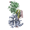
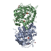
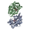
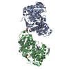
| Title | Crystal structure of the Escherichia coli nucleosidase PpnN (apo form) | ||||||
 Components Components | Pyrimidine/purine nucleotide 5'-monophosphate nucleosidase | ||||||
 Keywords Keywords | HYDROLASE / YgdH / PpnN / allosteric enzyme / nucleotide metabolism / stringent response / antibiotic tolerance / persistence / fluoroquinolone | ||||||
| Function / homology |  Function and homology information Function and homology informationinosinate nucleosidase activity / pyrimidine-5'-nucleotide nucleosidase / pyrimidine-5'-nucleotide nucleosidase activity / AMP nucleosidase / AMP nucleosidase activity / guanosine tetraphosphate binding / Hydrolases; Glycosylases; Hydrolysing N-glycosyl compounds / protein homotetramerization / protein-containing complex / identical protein binding / cytosol Similarity search - Function | ||||||
| Biological species |  | ||||||
| Method |  X-RAY DIFFRACTION / SYNCHROTRON / MOLECULAR REPLACEMENT / Resolution: 2.48 Å X-RAY DIFFRACTION / SYNCHROTRON / MOLECULAR REPLACEMENT / Resolution: 2.48 Å | ||||||
 Authors Authors | Zhang, Y. / Baerentsen, R.L. / Gerdes, K. / Brodersen, D.E. | ||||||
| Funding support |  Denmark, 1items Denmark, 1items
| ||||||
 Citation Citation | Journal: Mol.Cell / Year: 2019 Title: (p)ppGpp Regulates a Bacterial Nucleosidase by an Allosteric Two-Domain Switch. Authors: Zhang, Y.E. / Baerentsen, R.L. / Fuhrer, T. / Sauer, U. / Gerdes, K. / Brodersen, D.E. | ||||||
| History |
|
- Structure visualization
| Structure viewer | Molecule: MolmilJmol/JSmol |
|---|
- Downloads & links
-Download
| PDBx/mmCIF format | 6gfl.cif.gz | 361.1 KB | Display | PDBx/mmCIF format |
|---|---|---|---|---|
| PDB format | pdb6gfl.ent.gz | 294 KB | Display | PDB format |
| PDBx/mmJSON format | 6gfl.json.gz | Tree view | PDBx/mmJSON format | |
| Others |  Other downloads Other downloads |
-Validation report
| Arichive directory | https://data.pdbj.org/pub/pdb/validation_reports/gf/6gflftp://data.pdbj.org/pub/pdb/validation_reports/gf/6gfl | HTTPS FTP |
|---|
-Related structure data
| Related structure data |  6gfmC  2pmbS C: citing same article ( S: Starting model for refinement |
|---|---|
| Similar structure data |


-Links
 PDBj
PDBj- Assembly
| Deposited unit | 
| ||||||||||||
|---|---|---|---|---|---|---|---|---|---|---|---|---|---|
| 1 | 
| ||||||||||||
| Unit cell |
|
-Components
| #1: Protein | Mass: 53244.168 Da / Num. of mol.: 2 Source method: isolated from a genetically manipulated source Source: (gene. exp.) References: UniProt: P0ADR8, Hydrolases; Glycosylases; Hydrolysing N-glycosyl compounds, pyrimidine-5'-nucleotide nucleosidase, AMP nucleosidase #2: Water | ChemComp-HOH / |  Mass: 18.015 Da / Num. of mol.: 161 / Source method: isolated from a natural source / Formula: H2O Mass: 18.015 Da / Num. of mol.: 161 / Source method: isolated from a natural source / Formula: H2OHas protein modification | N | |
|---|
-Experimental details
-Experiment
| Experiment | Method: X-RAY DIFFRACTION / Number of used crystals: 1 |
|---|
- Sample preparation
| Crystal | Density Matthews: 2.86 Å3/Da / Density % sol: 57.08 % |
|---|---|
| Crystal grow | Temperature: 293.15 K / Method: vapor diffusion, sitting drop / pH: 7.5 Details: 0.1M HEPES pH 7.5 36% v/v PEG200 5 mg/ml protein concentration |
-Data collection
| Diffraction | Mean temperature: 100 K |
|---|---|
| Diffraction source | Source: SYNCHROTRON / Site: Diamond  / Beamline: I24 / Wavelength: 0.9686 Å / Beamline: I24 / Wavelength: 0.9686 Å |
| Detector | Type: DECTRIS PILATUS3 6M / Detector: PIXEL / Date: Oct 30, 2017 Details: Oxford Danfysik/SESO Two stage demagnification using two K-B pairs of bimorph type mirrors |
| Radiation | Monochromator: ACCEL Fixed exit Double Crystal / Protocol: SINGLE WAVELENGTH / Monochromatic (M) / Laue (L): M / Scattering type: x-ray |
| Radiation wavelength | Wavelength: 0.9686 Å / Relative weight: 1 |
| Reflection | Resolution: 2.48→49.08 Å / Num. obs: 38638 / % possible obs: 99.92 % / Redundancy: 13.4 % / Biso Wilson estimate: 65.26 Å2 / CC1/2: 0.99 / Rmerge(I) obs: 0.13 / Rpim(I) all: 0.03 / Rrim(I) all: 0.13 / Net I/σ(I): 12.97 |
| Reflection shell | Resolution: 2.48→2.57 Å / Redundancy: 7.4 % / Rmerge(I) obs: 1.74 / Mean I/σ(I) obs: 1.15 / Num. unique obs: 3781 / CC1/2: 0.56 / Rpim(I) all: 0.68 / Rrim(I) all: 1.87 / % possible all: 99.95 |
- Processing
| Software |
| ||||||||||||||||
|---|---|---|---|---|---|---|---|---|---|---|---|---|---|---|---|---|---|
| Refinement | Method to determine structure: MOLECULAR REPLACEMENT Starting model: 2PMB Resolution: 2.48→49.08 Å / Cross valid method: FREE R-VALUE
| ||||||||||||||||
| Refinement step | Cycle: LAST / Resolution: 2.48→49.08 Å
|
