 Movie
Movie Controller

[English] 日本語
 Yorodumi
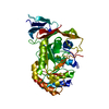
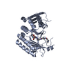
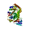
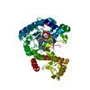
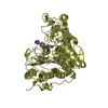
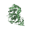
- PDB-6gdf: DIHYDROOROTASE FROM AQUIFEX AEOLICUS UNDER 600 BAR OF HYDROSTATIC... -
+ Open data

- Basic information
| Entry | Database: PDB / ID: 6gdf | ||||||
|---|---|---|---|---|---|---|---|
| Title | DIHYDROOROTASE FROM AQUIFEX AEOLICUS UNDER 600 BAR OF HYDROSTATIC PRESSURE | ||||||
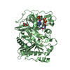
 Components Components | Dihydroorotase | ||||||
 Keywords Keywords | HYDROLASE / Dihydroorotase / Aquifex aeolicus / at 600 bar hydrostatic pressure / room temperature | ||||||
| Function / homology |  Function and homology information Function and homology informationallantoinase activity / dihydroorotase / purine nucleobase catabolic process / dihydroorotase activity / 'de novo' UMP biosynthetic process / zinc ion binding / cytoplasm Similarity search - Function | ||||||
| Biological species |   Aquifex aeolicus (bacteria) Aquifex aeolicus (bacteria) | ||||||
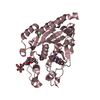
| Method |  X-RAY DIFFRACTION / SYNCHROTRON / MOLECULAR REPLACEMENT / Resolution: 2.5 Å X-RAY DIFFRACTION / SYNCHROTRON / MOLECULAR REPLACEMENT / Resolution: 2.5 Å | ||||||
 Authors Authors | Prange, T. / Girard, E. / Herve, G. / Evans, D.R. | ||||||
 Citation Citation | Journal: Febs J. / Year: 2019 Title: Pressure-induced activation of latent dihydroorotase from Aquifex aeolicus as revealed by high pressure protein crystallography. Authors: Prange, T. / Girard, E. / Fourme, R. / Dhaussy, A.C. / Edwards, B. / Vaishnav, A. / Patel, C. / Guy-Evans, H. / Herve, G. / Evans, D.R. #1: Journal: J. Biol. Chem. / Year: 2017 Title: Activation of latent dihydroorotase from aquifex aeolicus by pressure Authors: Herve, G. / Guy-Evans, H. / Fernado, R. / Patel, C. / Hachem, F. / Evans, D.R. | ||||||
| History |
|
- Structure visualization
| Structure viewer | Molecule: MolmilJmol/JSmol |
|---|
- Downloads & links
-Download
| PDBx/mmCIF format | 6gdf.cif.gz | 86.6 KB | Display | PDBx/mmCIF format |
|---|---|---|---|---|
| PDB format | pdb6gdf.ent.gz | 65.1 KB | Display | PDB format |
| PDBx/mmJSON format | 6gdf.json.gz | Tree view | PDBx/mmJSON format | |
| Others |  Other downloads Other downloads |
-Validation report
| Arichive directory | https://data.pdbj.org/pub/pdb/validation_reports/gd/6gdfftp://data.pdbj.org/pub/pdb/validation_reports/gd/6gdf | HTTPS FTP |
|---|
-Related structure data
| Related structure data |  6gddC  6gdeC  1xrfS S: Starting model for refinement C: citing same article ( |
|---|---|
| Similar structure data |


-Links
 PDBj
PDBj

- Assembly
| Deposited unit | 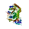
| ||||||||
|---|---|---|---|---|---|---|---|---|---|
| 1 |
| ||||||||
| Unit cell |
|
-Components
| #1: Protein | Mass: 46626.922 Da / Num. of mol.: 1 Source method: isolated from a genetically manipulated source Source: (gene. exp.) Aquifex aeolicus (bacteria) / Strain: VF5 / Gene: pyrC, aq_806 / Production host: |
|---|---|
| #2: Chemical | ChemComp-ZN /   Mass: 65.409 Da / Num. of mol.: 1 / Source method: obtained synthetically / Formula: Zn Mass: 65.409 Da / Num. of mol.: 1 / Source method: obtained synthetically / Formula: Zn |
| #3: Chemical | ChemComp-SO4 /   Mass: 96.063 Da / Num. of mol.: 1 / Source method: obtained synthetically / Formula: SO4 Mass: 96.063 Da / Num. of mol.: 1 / Source method: obtained synthetically / Formula: SO4 |
| #4: Water | ChemComp-HOH /  Mass: 18.015 Da / Num. of mol.: 87 / Source method: isolated from a natural source / Formula: H2O Mass: 18.015 Da / Num. of mol.: 87 / Source method: isolated from a natural source / Formula: H2O |
-Experimental details
-Experiment
| Experiment | Method: X-RAY DIFFRACTION / Number of used crystals: 1 |
|---|
- Sample preparation
| Crystal | Density Matthews: 3.28 Å3/Da / Density % sol: 62 % / Description: Prism, 0.2 mm |
|---|---|
| Crystal grow | Temperature: 298 K / Method: vapor diffusion, hanging drop / pH: 4.6 Details: ETHANOL (12%), PEG 400 (4%), ACETATE, PH 4.6 (0.1 M) |
-Data collection
| Diffraction | Mean temperature: 298 K |
|---|---|
| Diffraction source | Source: SYNCHROTRON / Site: ESRF  / Beamline: ID27 / Wavelength: 0.3738 Å / Beamline: ID27 / Wavelength: 0.3738 Å |
| Detector | Type: MARRESEARCH / Detector: CCD / Date: May 22, 2015 |
| Radiation | Protocol: SINGLE WAVELENGTH / Monochromatic (M) / Laue (L): M / Scattering type: x-ray |
| Radiation wavelength | Wavelength: 0.3738 Å / Relative weight: 1 |
| Reflection | Resolution: 2.5→45.42 Å / Num. obs: 14277 / % possible obs: 76.8 % / Redundancy: 4.7 % / Biso Wilson estimate: 46.1 Å2 / Rmerge(I) obs: 0.135 / Net I/σ(I): 8.3 |
| Reflection shell | Resolution: 2.5→2.64 Å / Redundancy: 4.7 % / Rmerge(I) obs: 0.833 / Mean I/σ(I) obs: 1.7 / % possible all: 78.3 |
- Processing
| Software |
| ||||||||||||||||||||||||||||||||||||||||||||||||||||||||||||||||||||||||||||||||||||||||||||||||||||||||||||||||||||||||||||||||||||||||||||||||||||||||||||||||||||||||||||||||||||||
|---|---|---|---|---|---|---|---|---|---|---|---|---|---|---|---|---|---|---|---|---|---|---|---|---|---|---|---|---|---|---|---|---|---|---|---|---|---|---|---|---|---|---|---|---|---|---|---|---|---|---|---|---|---|---|---|---|---|---|---|---|---|---|---|---|---|---|---|---|---|---|---|---|---|---|---|---|---|---|---|---|---|---|---|---|---|---|---|---|---|---|---|---|---|---|---|---|---|---|---|---|---|---|---|---|---|---|---|---|---|---|---|---|---|---|---|---|---|---|---|---|---|---|---|---|---|---|---|---|---|---|---|---|---|---|---|---|---|---|---|---|---|---|---|---|---|---|---|---|---|---|---|---|---|---|---|---|---|---|---|---|---|---|---|---|---|---|---|---|---|---|---|---|---|---|---|---|---|---|---|---|---|---|---|
| Refinement | Method to determine structure: MOLECULAR REPLACEMENT Starting model: 1XRF Resolution: 2.5→45.42 Å / Cor.coef. Fo:Fc: 0.958 / SU B: 6.304 / SU ML: 0.128 / Cross valid method: NONE / ESU R: 0.717 / Stereochemistry target values: MAXIMUM LIKELIHOOD
| ||||||||||||||||||||||||||||||||||||||||||||||||||||||||||||||||||||||||||||||||||||||||||||||||||||||||||||||||||||||||||||||||||||||||||||||||||||||||||||||||||||||||||||||||||||||
| Solvent computation | Ion probe radii: 0.8 Å / Shrinkage radii: 0.8 Å / VDW probe radii: 1.2 Å / Solvent model: MASK | ||||||||||||||||||||||||||||||||||||||||||||||||||||||||||||||||||||||||||||||||||||||||||||||||||||||||||||||||||||||||||||||||||||||||||||||||||||||||||||||||||||||||||||||||||||||
| Displacement parameters | Biso mean: 45.46 Å2
| ||||||||||||||||||||||||||||||||||||||||||||||||||||||||||||||||||||||||||||||||||||||||||||||||||||||||||||||||||||||||||||||||||||||||||||||||||||||||||||||||||||||||||||||||||||||
| Refinement step | Cycle: LAST / Resolution: 2.5→45.42 Å
| ||||||||||||||||||||||||||||||||||||||||||||||||||||||||||||||||||||||||||||||||||||||||||||||||||||||||||||||||||||||||||||||||||||||||||||||||||||||||||||||||||||||||||||||||||||||
| Refine LS restraints |
|