Mass: 18.015 Da / Num. of mol.: 68 / Source method: isolated from a natural source / Formula: H2O
-
Experimental details
-
Experiment
Experiment
Method: X-RAY DIFFRACTION / Number of used crystals: 1
-
Sample preparation
Crystal
Density Matthews: 3.11 Å3/Da / Density % sol: 60.48 %
Crystal grow
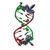
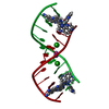
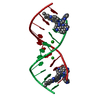
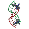
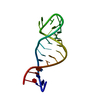
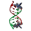
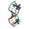
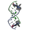
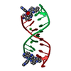
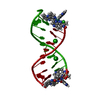
Temperature: 291 K / Method: vapor diffusion, sitting drop / pH: 7 Details: Crystallisation was observed in a number of conditions from the Nucleic Acid Mini-Screen from Hampton Research, where the best diffracting example came from a 8uL drop containing; 125 uM ...Details: Crystallisation was observed in a number of conditions from the Nucleic Acid Mini-Screen from Hampton Research, where the best diffracting example came from a 8uL drop containing; 125 uM d(CCGGACCCGG/CCGGGTCCGG)2, 750 uM rac-[Ru(TAP)2(11,12-CN2-dppz)]2+, 7.5% v/v MPD, 30 mM pH 7 sodium cacodylate, 9 mM spermine tetrahydrochloride, 60 mM KCl and 15 mM BaCl2; all equilibrated against 1 mL of 35% v/v MPD. Orange/red rods grew following roughly 3 weeks of incubation at 291 K.
In the structure databanks used in Yorodumi, some data are registered as the other names, "COVID-19 virus" and "2019-nCoV". Here are the details of the virus and the list of structure data.
Jan 31, 2019. EMDB accession codes are about to change! (news from PDBe EMDB page)
EMDB accession codes are about to change! (news from PDBe EMDB page)
The allocation of 4 digits for EMDB accession codes will soon come to an end. Whilst these codes will remain in use, new EMDB accession codes will include an additional digit and will expand incrementally as the available range of codes is exhausted. The current 4-digit format prefixed with “EMD-” (i.e. EMD-XXXX) will advance to a 5-digit format (i.e. EMD-XXXXX), and so on. It is currently estimated that the 4-digit codes will be depleted around Spring 2019, at which point the 5-digit format will come into force.
The EM Navigator/Yorodumi systems omit the EMD- prefix.
Related info.:Q: What is EMD? / ID/Accession-code notation in Yorodumi/EM Navigator
Yorodumi is a browser for structure data from EMDB, PDB, SASBDB, etc.
This page is also the successor to EM Navigator detail page, and also detail information page/front-end page for Omokage search.
The word "yorodu" (or yorozu) is an old Japanese word meaning "ten thousand". "mi" (miru) is to see.
Related info.:EMDB / PDB / SASBDB / Comparison of 3 databanks / Yorodumi Search / Aug 31, 2016. New EM Navigator & Yorodumi / Yorodumi Papers / Jmol/JSmol / Function and homology information / Changes in new EM Navigator and Yorodumi
 Movie
Movie Controller


 Open data
 Basic information
 Components
 Keywords
 Function and homology information
 X-RAY DIFFRACTION /
  Authors
 United Kingdom, 3items
  Citation
 Structure visualization
 Downloads & links
 Other downloads
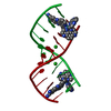


 PDBj
PDBj

 Assembly
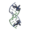

 Mass: 137.327 Da / Num. of mol.: 2 / Source method: obtained synthetically / Formula: Ba
 Mass: 797.751 Da / Num. of mol.: 2 / Source method: obtained synthetically / Formula: C40H20N14Ru
 Mass: 18.015 Da / Num. of mol.: 68 / Source method: isolated from a natural source / Formula: H2O
 Sample preparation
 Processing