 Movie
Movie Controller


+ Open data

- Basic information
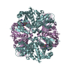
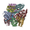
| Entry | Database: PDB / ID: 6g42 | ||||||
|---|---|---|---|---|---|---|---|
| Title | Crystal structure of mavirus penton protein | ||||||
 Components Components | Minor capsid protein | ||||||
 Keywords Keywords | VIRAL PROTEIN / single jelly-roll / capsid protein / virus | ||||||
| Function / homology | Minor capsid protein Function and homology information Function and homology information | ||||||
| Biological species |  Cafeteriavirus-dependent mavirus Cafeteriavirus-dependent mavirus | ||||||
| Method |  X-RAY DIFFRACTION / SYNCHROTRON / MOLECULAR REPLACEMENT / Resolution: 2.7 Å X-RAY DIFFRACTION / SYNCHROTRON / MOLECULAR REPLACEMENT / Resolution: 2.7 Å | ||||||
 Authors Authors | Born, D. / Reuter, L. / Meinhart, A. / Reinstein, J. | ||||||
 Citation Citation | Journal: Proc. Natl. Acad. Sci. U.S.A. / Year: 2018 Title: Capsid protein structure, self-assembly, and processing reveal morphogenesis of the marine virophage mavirus. Authors: Born, D. / Reuter, L. / Mersdorf, U. / Mueller, M. / Fischer, M.G. / Meinhart, A. / Reinstein, J. | ||||||
| History |
|
- Structure visualization
| Structure viewer | Molecule: MolmilJmol/JSmol |
|---|
- Downloads & links
-Download
| PDBx/mmCIF format | 6g42.cif.gz | 308.3 KB | Display | PDBx/mmCIF format |
|---|---|---|---|---|
| PDB format | pdb6g42.ent.gz | 251.5 KB | Display | PDB format |
| PDBx/mmJSON format | 6g42.json.gz | Tree view | PDBx/mmJSON format | |
| Others |  Other downloads Other downloads |
-Validation report
| Arichive directory | https://data.pdbj.org/pub/pdb/validation_reports/g4/6g42ftp://data.pdbj.org/pub/pdb/validation_reports/g4/6g42 | HTTPS FTP |
|---|
-Related structure data
| Related structure data | 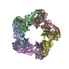 6g41SC  6g43C  6g44C  6g45C S: Starting model for refinement C: citing same article ( |
|---|---|
| Similar structure data |


-Links
 PDBj
PDBj- Assembly
| Deposited unit | 
| ||||||||||||||||||||||||||||||||||||||||||||||||||||||||||||||||||||||||||||||||||||||||||||||||||||||||||||||||||||||||||||||||||||||||||||||||||||||||||||
|---|---|---|---|---|---|---|---|---|---|---|---|---|---|---|---|---|---|---|---|---|---|---|---|---|---|---|---|---|---|---|---|---|---|---|---|---|---|---|---|---|---|---|---|---|---|---|---|---|---|---|---|---|---|---|---|---|---|---|---|---|---|---|---|---|---|---|---|---|---|---|---|---|---|---|---|---|---|---|---|---|---|---|---|---|---|---|---|---|---|---|---|---|---|---|---|---|---|---|---|---|---|---|---|---|---|---|---|---|---|---|---|---|---|---|---|---|---|---|---|---|---|---|---|---|---|---|---|---|---|---|---|---|---|---|---|---|---|---|---|---|---|---|---|---|---|---|---|---|---|---|---|---|---|---|---|---|---|
| 1 |
| ||||||||||||||||||||||||||||||||||||||||||||||||||||||||||||||||||||||||||||||||||||||||||||||||||||||||||||||||||||||||||||||||||||||||||||||||||||||||||||
| Unit cell |
| ||||||||||||||||||||||||||||||||||||||||||||||||||||||||||||||||||||||||||||||||||||||||||||||||||||||||||||||||||||||||||||||||||||||||||||||||||||||||||||
| Noncrystallographic symmetry (NCS) | NCS domain:
NCS domain segments: Ens-ID: 1 / Refine code: 4
|