 Movie
Movie Controller

[English] 日本語
 Yorodumi
- PDB-6g0a: The crystal structure of the Pol2 catalytic domain of DNA polymer... -
+ Open data

- Basic information
| Entry | Database: PDB / ID: 6g0a | ||||||||||||
|---|---|---|---|---|---|---|---|---|---|---|---|---|---|
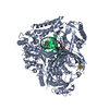
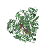
| Title | The crystal structure of the Pol2 catalytic domain of DNA polymerase epsilon carrying a P301R substitution. | ||||||||||||
 Components Components |
| ||||||||||||
 Keywords Keywords | DNA BINDING PROTEIN / DNA / Pol2 / PolE / Epsilon / P301R / cancer | ||||||||||||
| Function / homology |  Function and homology information Function and homology informationgene conversion / DNA replication initiation / epsilon DNA polymerase complex / SUMO binding / nucleotide-excision repair, DNA gap filling / Activation of the pre-replicative complex / DNA replication proofreading / Termination of translesion DNA synthesis / single-stranded DNA 3'-5' DNA exonuclease activity / mitotic DNA replication checkpoint signaling ...gene conversion / DNA replication initiation / epsilon DNA polymerase complex / SUMO binding / nucleotide-excision repair, DNA gap filling / Activation of the pre-replicative complex / DNA replication proofreading / Termination of translesion DNA synthesis / single-stranded DNA 3'-5' DNA exonuclease activity / mitotic DNA replication checkpoint signaling / mitotic intra-S DNA damage checkpoint signaling / Hydrolases; Acting on ester bonds; Exodeoxyribonucleases producing 5'-phosphomonoesters / mitotic sister chromatid cohesion / leading strand elongation / nuclear replication fork / Dual incision in TC-NER / error-prone translesion synthesis / base-excision repair, gap-filling / replication fork / base-excision repair / double-strand break repair via nonhomologous end joining / DNA-templated DNA replication / double-strand break repair / mitotic cell cycle / single-stranded DNA binding / 4 iron, 4 sulfur cluster binding / double-stranded DNA binding / DNA-directed DNA polymerase / DNA-directed DNA polymerase activity / nucleotide binding / mRNA binding / DNA binding / zinc ion binding / nucleus Similarity search - Function | ||||||||||||
| Biological species |  synthetic construct (others) | ||||||||||||
| Method |  X-RAY DIFFRACTION / SYNCHROTRON / MOLECULAR REPLACEMENT / Resolution: 2.62 Å X-RAY DIFFRACTION / SYNCHROTRON / MOLECULAR REPLACEMENT / Resolution: 2.62 Å | ||||||||||||
 Authors Authors | Parkash, V. / Johansson, E. | ||||||||||||
| Funding support |  Sweden, 3items Sweden, 3items
| ||||||||||||
 Citation Citation | Journal: Nat Commun / Year: 2019 Title: Structural consequence of the most frequently recurring cancer-associated substitution in DNA polymerase epsilon. Authors: Parkash, V. / Kulkarni, Y. / Ter Beek, J. / Shcherbakova, P.V. / Kamerlin, S.C.L. / Johansson, E. | ||||||||||||
| History |
|
- Structure visualization
| Structure viewer | Molecule: MolmilJmol/JSmol |
|---|
- Downloads & links
-Download
| PDBx/mmCIF format | 6g0a.cif.gz | 253.2 KB | Display | PDBx/mmCIF format |
|---|---|---|---|---|
| PDB format | pdb6g0a.ent.gz | 191.7 KB | Display | PDB format |
| PDBx/mmJSON format | 6g0a.json.gz | Tree view | PDBx/mmJSON format | |
| Others |  Other downloads Other downloads |
-Validation report
| Summary document | 6g0a_validation.pdf.gz | 822.9 KB | Display | wwPDB validaton report |
|---|---|---|---|---|
| Full document | 6g0a_full_validation.pdf.gz | 841.3 KB | Display | |
| Data in XML | 6g0a_validation.xml.gz | 40.9 KB | Display | |
| Data in CIF | 6g0a_validation.cif.gz | 55.7 KB | Display | |
| Arichive directory | https://data.pdbj.org/pub/pdb/validation_reports/g0/6g0aftp://data.pdbj.org/pub/pdb/validation_reports/g0/6g0a | HTTPS FTP |
-Related structure data
| Related structure data | 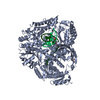 6fwkC 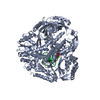 6i8aC  4m8oS C: citing same article ( S: Starting model for refinement |
|---|---|
| Similar structure data |


-Links
 PDBj
PDBj


- Assembly
| Deposited unit | 
| ||||||||
|---|---|---|---|---|---|---|---|---|---|
| 1 |
| ||||||||
| Unit cell |
|
-Components
-Protein , 1 types, 1 molecules A
| #1: Protein | Mass: 137235.672 Da / Num. of mol.: 1 / Mutation: P301R Source method: isolated from a genetically manipulated source Source: (gene. exp.) Gene: POL2, DUN2, YNL262W, N0825 / Production host:  |
|---|
-DNA chain , 2 types, 2 molecules PT
| #2: DNA chain | Mass: 3293.174 Da / Num. of mol.: 1 / Source method: obtained synthetically / Source: (synth.) synthetic construct (others) |
|---|---|
| #3: DNA chain | Mass: 4599.996 Da / Num. of mol.: 1 / Source method: obtained synthetically / Source: (synth.) synthetic construct (others) |
-Non-polymers , 4 types, 19 molecules 


| #4: Chemical | ChemComp-DTP /  Mass: 491.182 Da / Num. of mol.: 1 / Source method: obtained synthetically / Formula: C10H16N5O12P3 Mass: 491.182 Da / Num. of mol.: 1 / Source method: obtained synthetically / Formula: C10H16N5O12P3 | ||||
|---|---|---|---|---|---|
| #5: Chemical |  Mass: 40.078 Da / Num. of mol.: 2 / Source method: obtained synthetically / Formula: Ca Mass: 40.078 Da / Num. of mol.: 2 / Source method: obtained synthetically / Formula: Ca#6: Chemical | ChemComp-FE / |  Mass: 55.845 Da / Num. of mol.: 1 / Source method: obtained synthetically / Formula: Fe Mass: 55.845 Da / Num. of mol.: 1 / Source method: obtained synthetically / Formula: Fe#7: Water | ChemComp-HOH / | Mass: 18.015 Da / Num. of mol.: 15 / Source method: isolated from a natural source / Formula: H2O |
-Experimental details
-Experiment
| Experiment | Method: X-RAY DIFFRACTION / Number of used crystals: 1 |
|---|
- Sample preparation
| Crystal | Density Matthews: 2.68 Å3/Da / Density % sol: 54.17 % |
|---|---|
| Crystal grow | Temperature: 292 K / Method: vapor diffusion, hanging drop / pH: 6.5 / Details: 50mM MES pH 6.5, 150mM NaAc and 8% PEG20K / PH range: 6.5-7.0 |
-Data collection
| Diffraction | Mean temperature: 100 K | ||||||||||||||||||||||||||||||
|---|---|---|---|---|---|---|---|---|---|---|---|---|---|---|---|---|---|---|---|---|---|---|---|---|---|---|---|---|---|---|---|
| Diffraction source | Source: SYNCHROTRON / Site: ESRF  / Beamline: ID23-1 / Wavelength: 0.984 Å / Beamline: ID23-1 / Wavelength: 0.984 Å | ||||||||||||||||||||||||||||||
| Detector | Type: DECTRIS PILATUS3 6M / Detector: PIXEL / Date: Nov 4, 2017 | ||||||||||||||||||||||||||||||
| Radiation | Monochromator: Si (111) Silicon crystal / Protocol: SINGLE WAVELENGTH / Monochromatic (M) / Laue (L): M / Scattering type: x-ray | ||||||||||||||||||||||||||||||
| Radiation wavelength | Wavelength: 0.984 Å / Relative weight: 1 | ||||||||||||||||||||||||||||||
| Reflection | Resolution: 2.62→78.78 Å / Num. obs: 44773 / % possible obs: 98.7 % / Redundancy: 3 % / Biso Wilson estimate: 48.1 Å2 / CC1/2: 0.829 / Rmerge(I) obs: 0.128 / Rpim(I) all: 0.087 / Rrim(I) all: 0.156 / Net I/σ(I): 5.9 / Num. measured all: 136067 / Scaling rejects: 76 | ||||||||||||||||||||||||||||||
| Reflection shell | Diffraction-ID: 1
|
- Processing
| Software |
| |||||||||||||||||||||||||||||||||||||||||||||||||||||||||||||||||||||||||||||||||||||||||||||||||||||||||||||||||||||||
|---|---|---|---|---|---|---|---|---|---|---|---|---|---|---|---|---|---|---|---|---|---|---|---|---|---|---|---|---|---|---|---|---|---|---|---|---|---|---|---|---|---|---|---|---|---|---|---|---|---|---|---|---|---|---|---|---|---|---|---|---|---|---|---|---|---|---|---|---|---|---|---|---|---|---|---|---|---|---|---|---|---|---|---|---|---|---|---|---|---|---|---|---|---|---|---|---|---|---|---|---|---|---|---|---|---|---|---|---|---|---|---|---|---|---|---|---|---|---|---|---|
| Refinement | Method to determine structure: MOLECULAR REPLACEMENT Starting model: 4m8o Resolution: 2.62→19.977 Å / SU ML: 0.41 / Cross valid method: THROUGHOUT / σ(F): 1.34 / Phase error: 30.05 / Stereochemistry target values: ML
| |||||||||||||||||||||||||||||||||||||||||||||||||||||||||||||||||||||||||||||||||||||||||||||||||||||||||||||||||||||||
| Solvent computation | Shrinkage radii: 0.9 Å / VDW probe radii: 1.11 Å / Solvent model: FLAT BULK SOLVENT MODEL | |||||||||||||||||||||||||||||||||||||||||||||||||||||||||||||||||||||||||||||||||||||||||||||||||||||||||||||||||||||||
| Displacement parameters | Biso max: 112.3 Å2 / Biso mean: 53.9482 Å2 / Biso min: 20.86 Å2 | |||||||||||||||||||||||||||||||||||||||||||||||||||||||||||||||||||||||||||||||||||||||||||||||||||||||||||||||||||||||
| Refinement step | Cycle: final / Resolution: 2.62→19.977 Å
| |||||||||||||||||||||||||||||||||||||||||||||||||||||||||||||||||||||||||||||||||||||||||||||||||||||||||||||||||||||||
| LS refinement shell | Refine-ID: X-RAY DIFFRACTION / Rfactor Rfree error: 0 / Total num. of bins used: 16
|
