- PDB-6fwk: The crystal structure of Pol2CORE-M644G in complex with DNA and a... -
+
Open data
ID or keywords:
Loading...
-
Basic information
Entry
Database: PDB / ID: 6fwk
Title
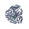
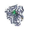
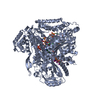
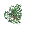
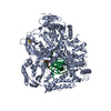
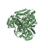
The crystal structure of Pol2CORE-M644G in complex with DNA and an incoming nucleotide
Components
DNA (5'-D(P*TP*AP*AP*CP*CP*GP*CP*GP*TP*TP*(DOC))-3')
DNA (5'-D(P*TP*CP*TP*TP*GP*AP*AP*CP*GP*CP*GP*GP*TP*TP*A)-3')
DNA polymerase epsilon catalytic subunit A
Keywords
DNA BINDING PROTEIN / Pol epsilon / M644G / DNA / complex
Function / homology
Function and homology information
gene conversion / DNA replication initiation / epsilon DNA polymerase complex / nucleotide-excision repair, DNA gap filling / SUMO binding / Activation of the pre-replicative complex / DNA replication proofreading / : / single-stranded DNA 3'-5' DNA exonuclease activity / mitotic DNA replication checkpoint signaling ...gene conversion / DNA replication initiation / epsilon DNA polymerase complex / nucleotide-excision repair, DNA gap filling / SUMO binding / Activation of the pre-replicative complex / DNA replication proofreading / : / single-stranded DNA 3'-5' DNA exonuclease activity / mitotic DNA replication checkpoint signaling / mitotic intra-S DNA damage checkpoint signaling / mitotic sister chromatid cohesion / Hydrolases; Acting on ester bonds; Exodeoxyribonucleases producing 5'-phosphomonoesters / leading strand elongation / nuclear replication fork / Dual incision in TC-NER / error-prone translesion synthesis / base-excision repair, gap-filling / replication fork / base-excision repair / DNA-templated DNA replication / double-strand break repair via nonhomologous end joining / double-strand break repair / mitotic cell cycle / single-stranded DNA binding / 4 iron, 4 sulfur cluster binding / double-stranded DNA binding / DNA-directed DNA polymerase / DNA-directed DNA polymerase activity / nucleotide binding / mRNA binding / DNA binding / zinc ion binding / nucleus Similarity search - Function
: / : / DNA polymerase epsilon catalytic subunit A, thumb domain / Zinc finger domain of DNA polymerase-epsilon / Zinc finger domain of DNA polymerase-epsilon / DNA polymerase epsilon, catalytic subunit A, C-terminal / DNA polymerase epsilon catalytic subunit / Domain of unknown function (DUF1744) / DUF1744 / DNA polymerase family B, thumb domain ...: / : / DNA polymerase epsilon catalytic subunit A, thumb domain / Zinc finger domain of DNA polymerase-epsilon / Zinc finger domain of DNA polymerase-epsilon / DNA polymerase epsilon, catalytic subunit A, C-terminal / DNA polymerase epsilon catalytic subunit / Domain of unknown function (DUF1744) / DUF1744 / DNA polymerase family B, thumb domain / DNA polymerase family B / DNA polymerase family B, exonuclease domain / DNA-directed DNA polymerase, family B, exonuclease domain / DNA polymerase, palm domain superfamily / DNA polymerase type-B family / DNA-directed DNA polymerase, family B / Ribonuclease H superfamily / Ribonuclease H-like superfamily / DNA/RNA polymerase superfamily Similarity search - Domain/homology
2'-DEOXYADENOSINE 5'-TRIPHOSPHATE / : / DNA / DNA (> 10) / DNA polymerase epsilon catalytic subunit A Similarity search - Component
A: DNA polymerase epsilon catalytic subunit A P: DNA (5'-D(P*TP*AP*AP*CP*CP*GP*CP*GP*TP*TP*(DOC))-3') T: DNA (5'-D(P*TP*CP*TP*TP*GP*AP*AP*CP*GP*CP*GP*GP*TP*TP*A)-3') B: DNA polymerase epsilon catalytic subunit A C: DNA (5'-D(P*TP*AP*AP*CP*CP*GP*CP*GP*TP*TP*(DOC))-3') D: DNA (5'-D(P*TP*CP*TP*TP*GP*AP*AP*CP*GP*CP*GP*GP*TP*TP*A)-3') hetero molecules
A: DNA polymerase epsilon catalytic subunit A P: DNA (5'-D(P*TP*AP*AP*CP*CP*GP*CP*GP*TP*TP*(DOC))-3') T: DNA (5'-D(P*TP*CP*TP*TP*GP*AP*AP*CP*GP*CP*GP*GP*TP*TP*A)-3') hetero molecules
B: DNA polymerase epsilon catalytic subunit A C: DNA (5'-D(P*TP*AP*AP*CP*CP*GP*CP*GP*TP*TP*(DOC))-3') D: DNA (5'-D(P*TP*CP*TP*TP*GP*AP*AP*CP*GP*CP*GP*GP*TP*TP*A)-3') hetero molecules
In the structure databanks used in Yorodumi, some data are registered as the other names, "COVID-19 virus" and "2019-nCoV". Here are the details of the virus and the list of structure data.
Jan 31, 2019. EMDB accession codes are about to change! (news from PDBe EMDB page)
EMDB accession codes are about to change! (news from PDBe EMDB page)
The allocation of 4 digits for EMDB accession codes will soon come to an end. Whilst these codes will remain in use, new EMDB accession codes will include an additional digit and will expand incrementally as the available range of codes is exhausted. The current 4-digit format prefixed with “EMD-” (i.e. EMD-XXXX) will advance to a 5-digit format (i.e. EMD-XXXXX), and so on. It is currently estimated that the 4-digit codes will be depleted around Spring 2019, at which point the 5-digit format will come into force.
The EM Navigator/Yorodumi systems omit the EMD- prefix.
Related info.:Q: What is EMD? / ID/Accession-code notation in Yorodumi/EM Navigator
Yorodumi is a browser for structure data from EMDB, PDB, SASBDB, etc.
This page is also the successor to EM Navigator detail page, and also detail information page/front-end page for Omokage search.
The word "yorodu" (or yorozu) is an old Japanese word meaning "ten thousand". "mi" (miru) is to see.
Related info.:EMDB / PDB / SASBDB / Comparison of 3 databanks / Yorodumi Search / Aug 31, 2016. New EM Navigator & Yorodumi / Yorodumi Papers / Jmol/JSmol / Function and homology information / Changes in new EM Navigator and Yorodumi
 Movie
Movie Controller

 Yorodumi
 Open data
 Basic information
 Components
 Keywords
 Function and homology information
 X-RAY DIFFRACTION /
  Authors
 Sweden, 3items
  Citation
 Structure visualization
 Downloads & links
 Other downloads
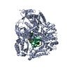


 PDBj
PDBj


 Assembly
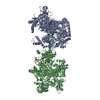


 Mass: 491.182 Da / Num. of mol.: 2 / Source method: obtained synthetically / Formula: C10H16N5O12P3
 Mass: 40.078 Da / Num. of mol.: 5 / Source method: obtained synthetically / Formula: Ca
 Mass: 55.845 Da / Num. of mol.: 2 / Source method: obtained synthetically / Formula: Fe
 Sample preparation
 / Beamline: ID23-1 / Wavelength: 0.97918 Å
 Processing