 Movie
Movie Controller

[English] 日本語
 Yorodumi
- PDB-6fyq: The crystal structure of a new transaminase from the marine bacte... -
+ Open data

- Basic information
| Entry | Database: PDB / ID: 6fyq | ||||||
|---|---|---|---|---|---|---|---|
| Title | The crystal structure of a new transaminase from the marine bacterium Virgibacillus | ||||||
 Components Components | amine transaminase | ||||||
 Keywords Keywords | TRANSFERASE / Omega-amino acid-pyruvate aminotransferase / Virgibacillus pantothenticus | ||||||
| Function / homology |  Function and homology information Function and homology informationbeta-alanine-pyruvate transaminase / beta-alanine:pyruvate transaminase activity / pyridoxal phosphate binding Similarity search - Function | ||||||
| Biological species |  Virgibacillus pantothenticus (bacteria) Virgibacillus pantothenticus (bacteria) | ||||||
| Method |  X-RAY DIFFRACTION / SYNCHROTRON / MOLECULAR REPLACEMENT / molecular replacement / Resolution: 2 Å X-RAY DIFFRACTION / SYNCHROTRON / MOLECULAR REPLACEMENT / molecular replacement / Resolution: 2 Å | ||||||
 Authors Authors | Gourlay, L.J. | ||||||
 Citation Citation | Journal: Sci Rep / Year: 2018 Title: Strategic single point mutation yields a solvent- and salt-stable transaminase from Virgibacillus sp. in soluble form. Authors: Guidi, B. / Planchestainer, M. / Contente, M.L. / Laurenzi, T. / Eberini, I. / Gourlay, L.J. / Romano, D. / Paradisi, F. / Molinari, F. | ||||||
| History |
|
- Structure visualization
| Structure viewer | Molecule: MolmilJmol/JSmol |
|---|
- Downloads & links
-Download
| PDBx/mmCIF format | 6fyq.cif.gz | 189.9 KB | Display | PDBx/mmCIF format |
|---|---|---|---|---|
| PDB format | pdb6fyq.ent.gz | 149.9 KB | Display | PDB format |
| PDBx/mmJSON format | 6fyq.json.gz | Tree view | PDBx/mmJSON format | |
| Others |  Other downloads Other downloads |
-Validation report
| Arichive directory | https://data.pdbj.org/pub/pdb/validation_reports/fy/6fyqftp://data.pdbj.org/pub/pdb/validation_reports/fy/6fyq | HTTPS FTP |
|---|
-Related structure data
| Related structure data | 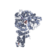 5tibS S: Starting model for refinement |
|---|---|
| Similar structure data |


-Links
 PDBj
PDBj- Assembly
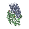
| Deposited unit | 
| ||||||||
|---|---|---|---|---|---|---|---|---|---|
| 1 | 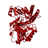
| ||||||||
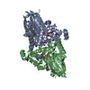
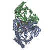
| Unit cell |
|
-Components
| #1: Protein | Mass: 53241.785 Da / Num. of mol.: 1 Source method: isolated from a genetically manipulated source Details: Contains an engineered point mutation T16F Source: (gene. exp.) Virgibacillus pantothenticus (bacteria)Strain: strain 21D / Plasmid: pET100D-TOPO / Production host: References: UniProt: A0A4P1LYG1*PLUS, beta-alanine-pyruvate transaminase | ||||
|---|---|---|---|---|---|
| #2: Chemical | ChemComp-PLP /   Mass: 247.142 Da / Num. of mol.: 1 / Source method: obtained synthetically / Formula: C8H10NO6P / Feature type: SUBJECT OF INVESTIGATION Mass: 247.142 Da / Num. of mol.: 1 / Source method: obtained synthetically / Formula: C8H10NO6P / Feature type: SUBJECT OF INVESTIGATION | ||||
| #3: Chemical |   Mass: 62.068 Da / Num. of mol.: 3 / Source method: obtained synthetically / Formula: C2H6O2 Mass: 62.068 Da / Num. of mol.: 3 / Source method: obtained synthetically / Formula: C2H6O2#4: Chemical | ChemComp-CL / |   Mass: 35.453 Da / Num. of mol.: 1 / Source method: obtained synthetically / Formula: Cl Mass: 35.453 Da / Num. of mol.: 1 / Source method: obtained synthetically / Formula: Cl#5: Water | ChemComp-HOH / |  Mass: 18.015 Da / Num. of mol.: 74 / Source method: isolated from a natural source / Formula: H2O Mass: 18.015 Da / Num. of mol.: 74 / Source method: isolated from a natural source / Formula: H2O |
-Experimental details
-Experiment
| Experiment | Method: X-RAY DIFFRACTION / Number of used crystals: 1 |
|---|
- Sample preparation
| Crystal | Density Matthews: 2.69 Å3/Da / Density % sol: 54.24 % |
|---|---|
| Crystal grow | Temperature: 293 K / Method: vapor diffusion, sitting drop / pH: 8.5 Details: Morpheus condition D10 (0.12 M Alcohols, 0.1 M Buffer System 3 pH 8.5, 50% (v/v) Precipitant Mix 2 (Molecular dimensions) |
-Data collection
| Diffraction | Mean temperature: 100 K |
|---|---|
| Diffraction source | Source: SYNCHROTRON / Site: ESRF  / Beamline: ID30B / Wavelength: 0.966 Å / Beamline: ID30B / Wavelength: 0.966 Å |
| Detector | Type: DECTRIS PILATUS 6M / Detector: PIXEL / Date: Nov 6, 2017 |
| Radiation | Protocol: SINGLE WAVELENGTH / Monochromatic (M) / Laue (L): M / Scattering type: x-ray |
| Radiation wavelength | Wavelength: 0.966 Å / Relative weight: 1 |
| Reflection | Resolution: 2→40 Å / Num. obs: 38942 / % possible obs: 99.6 % / Observed criterion σ(I): 2 / Redundancy: 3.8 % / Biso Wilson estimate: 41.25 Å2 / CC1/2: 0.999 / Rmerge(I) obs: 0.04 / Rpim(I) all: 0.023 / Rrim(I) all: 0.046 / Net I/σ(I): 17.3 |
| Reflection shell | Resolution: 2→2.11 Å / Redundancy: 3.9 % / Rmerge(I) obs: 0.393 / Mean I/σ(I) obs: 3.2 / Num. unique obs: 5629 / CC1/2: 0.888 / Rpim(I) all: 0.223 / Rrim(I) all: 0.454 / % possible all: 99.9 |
-Phasing
| Phasing | Method: molecular replacement |
|---|
- Processing
| Software |
| |||||||||||||||||||||||||||||||||||||||||||||||||||||||||||||||||||||||||||||||||||||||||||||||||||||||||
|---|---|---|---|---|---|---|---|---|---|---|---|---|---|---|---|---|---|---|---|---|---|---|---|---|---|---|---|---|---|---|---|---|---|---|---|---|---|---|---|---|---|---|---|---|---|---|---|---|---|---|---|---|---|---|---|---|---|---|---|---|---|---|---|---|---|---|---|---|---|---|---|---|---|---|---|---|---|---|---|---|---|---|---|---|---|---|---|---|---|---|---|---|---|---|---|---|---|---|---|---|---|---|---|---|---|---|
| Refinement | Method to determine structure: MOLECULAR REPLACEMENT Starting model: 5TIB Resolution: 2→39.86 Å / SU ML: 0.23 / Cross valid method: THROUGHOUT / σ(F): 1.34 / Phase error: 22.32
| |||||||||||||||||||||||||||||||||||||||||||||||||||||||||||||||||||||||||||||||||||||||||||||||||||||||||
| Solvent computation | Shrinkage radii: 0.9 Å / VDW probe radii: 1.11 Å | |||||||||||||||||||||||||||||||||||||||||||||||||||||||||||||||||||||||||||||||||||||||||||||||||||||||||
| Displacement parameters | Biso max: 104.52 Å2 / Biso mean: 49.98 Å2 / Biso min: 24.9 Å2 | |||||||||||||||||||||||||||||||||||||||||||||||||||||||||||||||||||||||||||||||||||||||||||||||||||||||||
| Refinement step | Cycle: final / Resolution: 2→39.86 Å
| |||||||||||||||||||||||||||||||||||||||||||||||||||||||||||||||||||||||||||||||||||||||||||||||||||||||||
| LS refinement shell | Refine-ID: X-RAY DIFFRACTION / Rfactor Rfree error: 0 / Total num. of bins used: 14
| |||||||||||||||||||||||||||||||||||||||||||||||||||||||||||||||||||||||||||||||||||||||||||||||||||||||||
| Refinement TLS params. | Method: refined / Refine-ID: X-RAY DIFFRACTION
| |||||||||||||||||||||||||||||||||||||||||||||||||||||||||||||||||||||||||||||||||||||||||||||||||||||||||
| Refinement TLS group |
|
