- PDB-6fws: Structure of DinG in complex with ssDNA and ADPBeF -
+
Open data
ID or keywords:
Loading...
-
Basic information
Entry
Database: PDB / ID: 6fws
Title
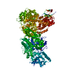
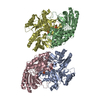
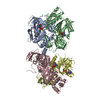
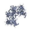
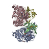
Structure of DinG in complex with ssDNA and ADPBeF
Components
ATP-dependent DNA helicase DinG
DNA (5'-D(*TP*TP*TP*TP*TP*TP*TP*TP*TP*T)-3')
DNA (5'-D(*TP*TP*TP*TP*TP*TP*TP*TP*TP*TP*T)-3')
Keywords
DNA BINDING PROTEIN / ATP / helicase / translocase / DNA binding
Function / homology
Function and homology information
catalytic activity, acting on a nucleic acid / DNA/RNA helicase activity / DNA 5'-3' helicase / SOS response / DNA helicase activity / 4 iron, 4 sulfur cluster binding / 5'-3' DNA helicase activity / DNA repair / ATP hydrolysis activity / DNA binding ...catalytic activity, acting on a nucleic acid / DNA/RNA helicase activity / DNA 5'-3' helicase / SOS response / DNA helicase activity / 4 iron, 4 sulfur cluster binding / 5'-3' DNA helicase activity / DNA repair / ATP hydrolysis activity / DNA binding / ATP binding / metal ion binding Similarity search - Function
A: ATP-dependent DNA helicase DinG C: DNA (5'-D(*TP*TP*TP*TP*TP*TP*TP*TP*TP*TP*T)-3') B: ATP-dependent DNA helicase DinG D: DNA (5'-D(*TP*TP*TP*TP*TP*TP*TP*TP*TP*T)-3') hetero molecules
In the structure databanks used in Yorodumi, some data are registered as the other names, "COVID-19 virus" and "2019-nCoV". Here are the details of the virus and the list of structure data.
Jan 31, 2019. EMDB accession codes are about to change! (news from PDBe EMDB page)
EMDB accession codes are about to change! (news from PDBe EMDB page)
The allocation of 4 digits for EMDB accession codes will soon come to an end. Whilst these codes will remain in use, new EMDB accession codes will include an additional digit and will expand incrementally as the available range of codes is exhausted. The current 4-digit format prefixed with “EMD-” (i.e. EMD-XXXX) will advance to a 5-digit format (i.e. EMD-XXXXX), and so on. It is currently estimated that the 4-digit codes will be depleted around Spring 2019, at which point the 5-digit format will come into force.
The EM Navigator/Yorodumi systems omit the EMD- prefix.
Related info.:Q: What is EMD? / ID/Accession-code notation in Yorodumi/EM Navigator
Yorodumi is a browser for structure data from EMDB, PDB, SASBDB, etc.
This page is also the successor to EM Navigator detail page, and also detail information page/front-end page for Omokage search.
The word "yorodu" (or yorozu) is an old Japanese word meaning "ten thousand". "mi" (miru) is to see.
Related info.:EMDB / PDB / SASBDB / Comparison of 3 databanks / Yorodumi Search / Aug 31, 2016. New EM Navigator & Yorodumi / Yorodumi Papers / Jmol/JSmol / Function and homology information / Changes in new EM Navigator and Yorodumi
 Movie
Movie Controller


 Open data
 Basic information
 Components
 Keywords
 Function and homology information
 X-RAY DIFFRACTION /
  Authors
 United Kingdom, 1items
  Citation
 Structure visualization
 Downloads & links
 Other downloads


 PDBj
PDBj


 Assembly


 Mass: 351.640 Da / Num. of mol.: 2 / Source method: isolated from a natural source / Formula: Fe4S4 / Feature type: SUBJECT OF INVESTIGATION
 Mass: 427.201 Da / Num. of mol.: 2 / Source method: obtained synthetically / Formula: C10H15N5O10P2 / Feature type: SUBJECT OF INVESTIGATION / Comment: ADP, energy-carrying molecule*YM
 Mass: 66.007 Da / Num. of mol.: 2 / Source method: obtained synthetically / Formula: BeF3 / Feature type: SUBJECT OF INVESTIGATION
 Mass: 24.305 Da / Num. of mol.: 2 / Source method: obtained synthetically / Formula: Mg / Feature type: SUBJECT OF INVESTIGATION
 Sample preparation
 Processing