 Movie
Movie Controller

[English] 日本語
 Yorodumi
- PDB-6fv4: The structure of N-acetyl-D-glucosamine-6-phosphate deacetylase D... -
+ Open data

- Basic information
| Entry | Database: PDB / ID: 6fv4 | |||||||||||||||||||||
|---|---|---|---|---|---|---|---|---|---|---|---|---|---|---|---|---|---|---|---|---|---|---|
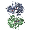
| Title | The structure of N-acetyl-D-glucosamine-6-phosphate deacetylase D267A mutant from Mycobacterium smegmatis in complex with N-acetyl-D-glucosamine-6-phosphate | |||||||||||||||||||||
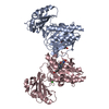
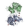
 Components Components | N-acetylglucosamine-6-phosphate deacetylase | |||||||||||||||||||||
 Keywords Keywords | HYDROLASE / N-acetyl-D-glucosamine-6-phosphate / mycobacteria carbohydrate metabolism | |||||||||||||||||||||
| Function / homology |  Function and homology information Function and homology informationN-acetylglucosamine-6-phosphate deacetylase / N-acetylglucosamine-6-phosphate deacetylase activity / N-acetylglucosamine catabolic process / metal ion binding Similarity search - Function | |||||||||||||||||||||
| Biological species |  Mycobacterium smegmatis (bacteria) Mycobacterium smegmatis (bacteria) | |||||||||||||||||||||
| Method |  X-RAY DIFFRACTION / SYNCHROTRON / MOLECULAR REPLACEMENT / Resolution: 1.974 Å X-RAY DIFFRACTION / SYNCHROTRON / MOLECULAR REPLACEMENT / Resolution: 1.974 Å | |||||||||||||||||||||
 Authors Authors | Ahangar, M.S. / Furze, C.M. / Guy, C.S. / Cooper, C. / Maskew, K.S. / Graham, B. / Cameron, A.D. / Fullam, E. | |||||||||||||||||||||
| Funding support |  United Kingdom, 6items United Kingdom, 6items
| |||||||||||||||||||||
 Citation Citation | Journal: J. Biol. Chem. / Year: 2018 Title: Structural and functional determination of homologs of theMycobacterium tuberculosis N-acetylglucosamine-6-phosphate deacetylase (NagA). Authors: Ahangar, M.S. / Furze, C.M. / Guy, C.S. / Cooper, C. / Maskew, K.S. / Graham, B. / Cameron, A.D. / Fullam, E. | |||||||||||||||||||||
| History |
|
- Structure visualization
| Structure viewer | Molecule: MolmilJmol/JSmol |
|---|
- Downloads & links
-Download
| PDBx/mmCIF format | 6fv4.cif.gz | 165.9 KB | Display | PDBx/mmCIF format |
|---|---|---|---|---|
| PDB format | pdb6fv4.ent.gz | 127.9 KB | Display | PDB format |
| PDBx/mmJSON format | 6fv4.json.gz | Tree view | PDBx/mmJSON format | |
| Others |  Other downloads Other downloads |
-Validation report
| Arichive directory | https://data.pdbj.org/pub/pdb/validation_reports/fv/6fv4ftp://data.pdbj.org/pub/pdb/validation_reports/fv/6fv4 | HTTPS FTP |
|---|
-Related structure data
| Related structure data |  6fv3SC S: Starting model for refinement C: citing same article ( |
|---|---|
| Similar structure data |


-Links
 PDBj
PDBj


- Assembly
| Deposited unit | 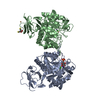
| ||||||||
|---|---|---|---|---|---|---|---|---|---|
| 1 |
| ||||||||
| Unit cell |
| ||||||||
| Components on special symmetry positions |
|
-Components
-Protein / Sugars , 2 types, 3 molecules AB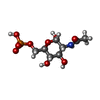
| #1: Protein | Mass: 41272.738 Da / Num. of mol.: 2 / Mutation: D267A Source method: isolated from a genetically manipulated source Source: (gene. exp.) Mycobacterium smegmatis (strain ATCC 700084 / mc(2)155) (bacteria)Strain: ATCC 700084 / mc(2)155 / Gene: nagA, MSMEG_2119 / Production host: References: UniProt: A0QU89, N-acetylglucosamine-6-phosphate deacetylase #3: Sugar | ChemComp-16G / |  Type: D-saccharide, alpha linking / Mass: 301.188 Da / Num. of mol.: 1 Type: D-saccharide, alpha linking / Mass: 301.188 Da / Num. of mol.: 1Source method: isolated from a genetically manipulated source Formula: C8H16NO9P |
|---|
-Non-polymers , 5 types, 536 molecules 


| #2: Chemical |  Mass: 65.409 Da / Num. of mol.: 2 / Source method: obtained synthetically / Formula: Zn Mass: 65.409 Da / Num. of mol.: 2 / Source method: obtained synthetically / Formula: Zn#4: Chemical | ChemComp-CD /  Mass: 112.411 Da / Num. of mol.: 4 / Source method: obtained synthetically / Formula: Cd Mass: 112.411 Da / Num. of mol.: 4 / Source method: obtained synthetically / Formula: Cd#5: Chemical |  Mass: 35.453 Da / Num. of mol.: 2 / Source method: obtained synthetically / Formula: Cl Mass: 35.453 Da / Num. of mol.: 2 / Source method: obtained synthetically / Formula: Cl#6: Chemical | ChemComp-DTT / |  Mass: 154.251 Da / Num. of mol.: 1 / Source method: obtained synthetically / Formula: C4H10O2S2 Mass: 154.251 Da / Num. of mol.: 1 / Source method: obtained synthetically / Formula: C4H10O2S2#7: Water | ChemComp-HOH / | Mass: 18.015 Da / Num. of mol.: 527 / Source method: isolated from a natural source / Formula: H2O |
|---|
-Experimental details
-Experiment
| Experiment | Method: X-RAY DIFFRACTION / Number of used crystals: 1 |
|---|
- Sample preparation
| Crystal | Density Matthews: 2.51 Å3/Da / Density % sol: 50.91 % |
|---|---|
| Crystal grow | Temperature: 295.15 K / Method: vapor diffusion Details: NagA crystals grew within a week at 295.15 K in 0.12 M monosaccharide mix (Morpheus, Molecular Dimensions), 0.1 M imidazole/MES pH 6.5, 20 % PEG 500 MME, 10 % w/v PEG 20000 with the addition ...Details: NagA crystals grew within a week at 295.15 K in 0.12 M monosaccharide mix (Morpheus, Molecular Dimensions), 0.1 M imidazole/MES pH 6.5, 20 % PEG 500 MME, 10 % w/v PEG 20000 with the addition of 10 mM CdCl2 additive. 5 mM GlcNAc6P was added and incubated at 277.14 K for 30 min before crystallization. |
-Data collection
| Diffraction | Mean temperature: 100 K |
|---|---|
| Diffraction source | Source: SYNCHROTRON / Site: Diamond / Beamline: I24 / Wavelength: 0.96863 Å |
| Detector | Type: DECTRIS PILATUS3 6M / Detector: PIXEL / Date: Jul 14, 2017 |
| Radiation | Protocol: SINGLE WAVELENGTH / Monochromatic (M) / Laue (L): M / Scattering type: x-ray |
| Radiation wavelength | Wavelength: 0.96863 Å / Relative weight: 1 |
| Reflection | Resolution: 1.974→80.141 Å / Num. obs: 55882 / % possible obs: 96.8 % / Redundancy: 6.5 % / CC1/2: 0.993 / Rmerge(I) obs: 0.128 / Rpim(I) all: 0.061 / Rrim(I) all: 0.152 / Net I/av σ(I): 8.5 / Net I/σ(I): 9.1 |
| Reflection shell | Resolution: 1.974→2.08 Å / Redundancy: 6.3 % / Rmerge(I) obs: 0.822 / Mean I/σ(I) obs: 2.7 / Num. unique obs: 8181 / CC1/2: 0.841 / Rpim(I) all: 0.412 / Rrim(I) all: 0.987 / % possible all: 98.1 |
- Processing
| Software |
| |||||||||||||||||||||||||||||||||||||||||||||||||||||||||||||||||||||||||||||||||||||||||||||||||||||||||||||||||||||||||||||||||||||||||||||||||||
|---|---|---|---|---|---|---|---|---|---|---|---|---|---|---|---|---|---|---|---|---|---|---|---|---|---|---|---|---|---|---|---|---|---|---|---|---|---|---|---|---|---|---|---|---|---|---|---|---|---|---|---|---|---|---|---|---|---|---|---|---|---|---|---|---|---|---|---|---|---|---|---|---|---|---|---|---|---|---|---|---|---|---|---|---|---|---|---|---|---|---|---|---|---|---|---|---|---|---|---|---|---|---|---|---|---|---|---|---|---|---|---|---|---|---|---|---|---|---|---|---|---|---|---|---|---|---|---|---|---|---|---|---|---|---|---|---|---|---|---|---|---|---|---|---|---|---|---|---|
| Refinement | Method to determine structure: MOLECULAR REPLACEMENT Starting model: 6FV3 Resolution: 1.974→54.56 Å / SU ML: 0.19 / Cross valid method: FREE R-VALUE / σ(F): 1.35 / Phase error: 26.66 / Stereochemistry target values: ML
| |||||||||||||||||||||||||||||||||||||||||||||||||||||||||||||||||||||||||||||||||||||||||||||||||||||||||||||||||||||||||||||||||||||||||||||||||||
| Solvent computation | Shrinkage radii: 0.9 Å / VDW probe radii: 1.11 Å / Solvent model: FLAT BULK SOLVENT MODEL | |||||||||||||||||||||||||||||||||||||||||||||||||||||||||||||||||||||||||||||||||||||||||||||||||||||||||||||||||||||||||||||||||||||||||||||||||||
| Refinement step | Cycle: LAST / Resolution: 1.974→54.56 Å
| |||||||||||||||||||||||||||||||||||||||||||||||||||||||||||||||||||||||||||||||||||||||||||||||||||||||||||||||||||||||||||||||||||||||||||||||||||
| Refine LS restraints |
| |||||||||||||||||||||||||||||||||||||||||||||||||||||||||||||||||||||||||||||||||||||||||||||||||||||||||||||||||||||||||||||||||||||||||||||||||||
| LS refinement shell |
|
