 Movie
Movie Controller


+ Open data

- Basic information
| Entry | Database: PDB / ID: 6fkm | |||||||||
|---|---|---|---|---|---|---|---|---|---|---|
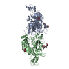
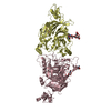
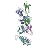
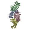
| Title | Drosophila Plexin A in complex with Semaphorin 1b | |||||||||
 Components Components |
| |||||||||
 Keywords Keywords | SIGNALING PROTEIN / semaphorin / plexin / sema domain / cell-cell signaling | |||||||||
| Function / homology |  Function and homology information Function and homology information: / Sema3A PAK dependent Axon repulsion / : / SEMA3A-Plexin repulsion signaling by inhibiting Integrin adhesion / guanylate cyclase activator activity / Sema4D induced cell migration and growth-cone collapse / Other semaphorin interactions / embryonic development via the syncytial blastoderm / photoreceptor cell axon guidance / semaphorin receptor binding ...: / Sema3A PAK dependent Axon repulsion / : / SEMA3A-Plexin repulsion signaling by inhibiting Integrin adhesion / guanylate cyclase activator activity / Sema4D induced cell migration and growth-cone collapse / Other semaphorin interactions / embryonic development via the syncytial blastoderm / photoreceptor cell axon guidance / semaphorin receptor binding / sensory neuron axon guidance / semaphorin receptor complex / chemorepellent activity / semaphorin receptor activity / axon midline choice point recognition / neural crest cell migration / negative chemotaxis / semaphorin-plexin signaling pathway / 14-3-3 protein binding / synapse assembly / regulation of cell migration / axon guidance / GTPase activator activity / heparin binding / Ras protein signal transduction / positive regulation of cell migration / plasma membrane / cytosol Similarity search - Function | |||||||||
| Biological species |  | |||||||||
| Method |  X-RAY DIFFRACTION / SYNCHROTRON / MOLECULAR REPLACEMENT / Resolution: 2.96 Å X-RAY DIFFRACTION / SYNCHROTRON / MOLECULAR REPLACEMENT / Resolution: 2.96 Å | |||||||||
 Authors Authors | Rozbesky, D. / Harlos, K. / Jones, E.Y. | |||||||||
| Funding support |  United Kingdom, 1items United Kingdom, 1items
| |||||||||
 Citation Citation | Journal: Embo J. / Year: 2020 Title: Structural basis of semaphorin-plexin cis interaction. Authors: Rozbesky, D. / Verhagen, M.G. / Karia, D. / Nagy, G.N. / Alvarez, L. / Robinson, R.A. / Harlos, K. / Padilla-Parra, S. / Pasterkamp, R.J. / Jones, E.Y. | |||||||||
| History |
|
- Structure visualization
| Structure viewer | Molecule: MolmilJmol/JSmol |
|---|
- Downloads & links
-Download
| PDBx/mmCIF format | 6fkm.cif.gz | 430.9 KB | Display | PDBx/mmCIF format |
|---|---|---|---|---|
| PDB format | pdb6fkm.ent.gz | 346.9 KB | Display | PDB format |
| PDBx/mmJSON format | 6fkm.json.gz | Tree view | PDBx/mmJSON format | |
| Others |  Other downloads Other downloads |
-Validation report
| Arichive directory | https://data.pdbj.org/pub/pdb/validation_reports/fk/6fkmftp://data.pdbj.org/pub/pdb/validation_reports/fk/6fkm | HTTPS FTP |
|---|
-Related structure data
| Related structure data |  6fknC 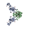 3okyS 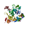 6fkkS S: Starting model for refinement C: citing same article ( |
|---|---|
| Similar structure data |


-Links
 PDBj
PDBj


- Assembly
| Deposited unit | 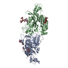
| ||||||||||
|---|---|---|---|---|---|---|---|---|---|---|---|
| 1 |
| ||||||||||
| Unit cell |
|
-Components
| #1: Protein | Mass: 78699.383 Da / Num. of mol.: 1 Source method: isolated from a genetically manipulated source Source: (gene. exp.) Gene: PlexA, BcDNA:GM05237, D-Plex A, Dmel\CG11081, DPlexA, lincRNA.927, plex, plex A, Plex1, plexA, PlexA1, CG11081, Dmel_CG11081 Variant: isoform A / Plasmid: pHLSec / Cell line (production host): HEK293T / Production host:  Homo sapiens (human) / References: UniProt: Q9V491 Homo sapiens (human) / References: UniProt: Q9V491 | ||||
|---|---|---|---|---|---|
| #2: Protein | Mass: 64212.426 Da / Num. of mol.: 1 Source method: isolated from a genetically manipulated source Source: (gene. exp.) Gene: Sema1b, Sema-1b, Sema-1b-RB, semaphorin-like, CG6446, Dmel_CG6446 Plasmid: pHLSec / Cell line (production host): HEK293T / Production host: Homo sapiens (human) / References: UniProt: Q7KK54 | ||||
| #3: Polysaccharide | Source method: isolated from a genetically manipulated source #4: Sugar | ChemComp-NAG /   Type: D-saccharide, beta linking / Mass: 221.208 Da / Num. of mol.: 4 Type: D-saccharide, beta linking / Mass: 221.208 Da / Num. of mol.: 4Source method: isolated from a genetically manipulated source Formula: C8H15NO6 Has protein modification | Y | |
-Experimental details
-Experiment
| Experiment | Method: X-RAY DIFFRACTION / Number of used crystals: 1 |
|---|
- Sample preparation
| Crystal | Density Matthews: 3.86 Å3/Da / Density % sol: 68.86 % |
|---|---|
| Crystal grow | Temperature: 293 K / Method: vapor diffusion, sitting drop / pH: 8 / Details: 0.1 M HEPES (pH 7.0) and 8% (w/v) PEG 8000 |
-Data collection
| Diffraction | Mean temperature: 100 K |
|---|---|
| Diffraction source | Source: SYNCHROTRON / Site: Diamond / Beamline: I24 / Wavelength: 1.072 Å |
| Detector | Type: DECTRIS PILATUS 6M / Detector: PIXEL / Date: Jul 3, 2016 |
| Radiation | Protocol: SINGLE WAVELENGTH / Monochromatic (M) / Laue (L): M / Scattering type: x-ray |
| Radiation wavelength | Wavelength: 1.072 Å / Relative weight: 1 |
| Reflection | Resolution: 2.96→76.864 Å / Num. obs: 31584 / % possible obs: 93.78 % / Redundancy: 4.6 % / Biso Wilson estimate: 69.89 Å2 / CC1/2: 0.99 / Rmerge(I) obs: 0.174 / Net I/σ(I): 8.86 |
| Reflection shell | Resolution: 2.96→3.07 Å / Rmerge(I) obs: 0.634 |
- Processing
| Software |
| ||||||||||||||||||||||||||||||||||||||||||||||||||||||||||||||||||||||||||||||||||||
|---|---|---|---|---|---|---|---|---|---|---|---|---|---|---|---|---|---|---|---|---|---|---|---|---|---|---|---|---|---|---|---|---|---|---|---|---|---|---|---|---|---|---|---|---|---|---|---|---|---|---|---|---|---|---|---|---|---|---|---|---|---|---|---|---|---|---|---|---|---|---|---|---|---|---|---|---|---|---|---|---|---|---|---|---|---|
| Refinement | Method to determine structure: MOLECULAR REPLACEMENT Starting model: 3OKY, 6FKK Resolution: 2.96→76.864 Å / SU ML: 0.44 / Cross valid method: THROUGHOUT / σ(F): 1.34 / Phase error: 26.8
| ||||||||||||||||||||||||||||||||||||||||||||||||||||||||||||||||||||||||||||||||||||
| Solvent computation | Shrinkage radii: 0.9 Å / VDW probe radii: 1.11 Å | ||||||||||||||||||||||||||||||||||||||||||||||||||||||||||||||||||||||||||||||||||||
| Refinement step | Cycle: LAST / Resolution: 2.96→76.864 Å
| ||||||||||||||||||||||||||||||||||||||||||||||||||||||||||||||||||||||||||||||||||||
| Refine LS restraints |
| ||||||||||||||||||||||||||||||||||||||||||||||||||||||||||||||||||||||||||||||||||||
| LS refinement shell |
| ||||||||||||||||||||||||||||||||||||||||||||||||||||||||||||||||||||||||||||||||||||
| Refinement TLS params. | Method: refined / Origin x: 43.7609 Å / Origin y: -9.6535 Å / Origin z: -38.6108 Å
| ||||||||||||||||||||||||||||||||||||||||||||||||||||||||||||||||||||||||||||||||||||
| Refinement TLS group | Selection details: all |
