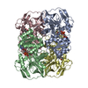
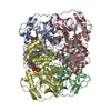
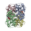
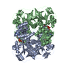
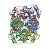
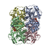
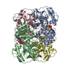
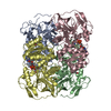
Entry Database : PDB / ID : 6fg0Title Crystal structure of R. ruber ADH-A, mutant Y54G, F43T, L119Y, F282W Alcohol dehydrogenase Keywords / / / / Function / homology Function Domain/homology Component
/ / / / / / / / / / / / / / / / / / / / / / / / / Biological species Rhodococcus sp. M8 (bacteria)Method / / / Resolution : 1.74 Å Authors Dobritzsch, D. / Maurer, D. / Hamnevik, E. / Enugala, T.R. / Widersten, M. Funding support Organization Grant number Country Swedish Research Council
Journal : Acs Catalysis / Year : 2018Title : Stereo- and Regioselectivity in Catalyzed Transformation of a 1,2-Disubstituted Vicinal Diol and the Corresponding Diketone by Wild Type and Laboratory Evolved Alcohol DehydrogenasesAuthors : Maurer, D. / Enugala, T.R. / Hamnevik, E. / Bauer, P. / Luking, M. / Hillier, H. / Kamerlin, S.C.L. / Dobritzsch, D. / Widersten, M. History Deposition Jan 9, 2018 Deposition site / Processing site Revision 1.0 Dec 19, 2018 Provider / Type Revision 1.1 Aug 7, 2019 Group / Database references / Category Item _citation.country / _citation.journal_abbrev ... _citation.country / _citation.journal_abbrev / _citation.journal_id_CSD / _citation.journal_id_ISSN / _citation.journal_volume / _citation.page_first / _citation.page_last / _citation.pdbx_database_id_DOI / _citation.title / _citation.year Revision 1.2 Jan 17, 2024 Group / Database references / Refinement descriptionCategory chem_comp_atom / chem_comp_bond ... chem_comp_atom / chem_comp_bond / database_2 / pdbx_initial_refinement_model / struct_ncs_dom_lim Item _database_2.pdbx_DOI / _database_2.pdbx_database_accession ... _database_2.pdbx_DOI / _database_2.pdbx_database_accession / _struct_ncs_dom_lim.beg_auth_comp_id / _struct_ncs_dom_lim.beg_label_asym_id / _struct_ncs_dom_lim.beg_label_comp_id / _struct_ncs_dom_lim.beg_label_seq_id / _struct_ncs_dom_lim.end_auth_comp_id / _struct_ncs_dom_lim.end_label_asym_id / _struct_ncs_dom_lim.end_label_comp_id / _struct_ncs_dom_lim.end_label_seq_id
Show all Show less
 Movie
Movie Controller

 Yorodumi
 Open data
 Basic information
 Components
 Keywords
 Function and homology information
 Rhodococcus sp. M8 (bacteria)
 X-RAY DIFFRACTION /
  Authors
 Sweden, 1items
  Citation
 Structure visualization
 Downloads & links
 Other downloads


 PDBj
PDBj

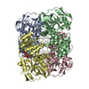
 Assembly
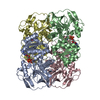

 Mass: 65.409 Da / Num. of mol.: 8 / Source method: obtained synthetically / Formula: Zn
 Mass: 663.425 Da / Num. of mol.: 4 / Source method: obtained synthetically / Formula: C21H27N7O14P2 / Comment: NAD*YM
 Mass: 18.015 Da / Num. of mol.: 1381 / Source method: isolated from a natural source / Formula: H2O
 Sample preparation
 / Beamline: I24 / Wavelength: 0.9686 Å
 Processing