 Movie
Movie Controller

[English] 日本語
 Yorodumi
- PDB-6fd6: Crystal Structure of Human APRT-Tyr105Phe variant in complex with... -
+ Open data

- Basic information
| Entry | Database: PDB / ID: 6fd6 | ||||||
|---|---|---|---|---|---|---|---|
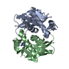
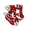
| Title | Crystal Structure of Human APRT-Tyr105Phe variant in complex with Adenine, PRPP and Mg2+, 30 days post crystallization (with AMP and PPi products fully generated) | ||||||
 Components Components | Adenine phosphoribosyltransferase | ||||||
 Keywords Keywords | TRANSFERASE / Rossman fold | ||||||
| Function / homology |  Function and homology information Function and homology informationDefective APRT disrupts adenine salvage / adenine binding / adenine salvage / adenine phosphoribosyltransferase / adenine phosphoribosyltransferase activity / Purine salvage / IMP salvage / AMP salvage / purine ribonucleoside salvage / AMP binding ...Defective APRT disrupts adenine salvage / adenine binding / adenine salvage / adenine phosphoribosyltransferase / adenine phosphoribosyltransferase activity / Purine salvage / IMP salvage / AMP salvage / purine ribonucleoside salvage / AMP binding / secretory granule lumen / Neutrophil degranulation / extracellular exosome / extracellular region / cytosol / cytoplasm Similarity search - Function | ||||||
| Biological species |  Homo sapiens (human) Homo sapiens (human) | ||||||
| Method |  X-RAY DIFFRACTION / SYNCHROTRON / MOLECULAR REPLACEMENT / Resolution: 1.8 Å X-RAY DIFFRACTION / SYNCHROTRON / MOLECULAR REPLACEMENT / Resolution: 1.8 Å | ||||||
| Model details | APO | ||||||
 Authors Authors | Nioche, P. / Huyet, J. / Ozeir, M. | ||||||
 Citation Citation | Journal: Cell Chem Biol / Year: 2018 Title: Structural Insights into the Forward and Reverse Enzymatic Reactions in Human Adenine Phosphoribosyltransferase. Authors: Huyet, J. / Ozeir, M. / Burgevin, M.C. / Pinson, B. / Chesney, F. / Remy, J.M. / Siddiqi, A.R. / Lupoli, R. / Pinon, G. / Saint-Marc, C. / Gibert, J.F. / Morales, R. / Ceballos-Picot, I. / ...Authors: Huyet, J. / Ozeir, M. / Burgevin, M.C. / Pinson, B. / Chesney, F. / Remy, J.M. / Siddiqi, A.R. / Lupoli, R. / Pinon, G. / Saint-Marc, C. / Gibert, J.F. / Morales, R. / Ceballos-Picot, I. / Barouki, R. / Daignan-Fornier, B. / Olivier-Bandini, A. / Auge, F. / Nioche, P. | ||||||
| History |
|
- Structure visualization
| Structure viewer | Molecule: MolmilJmol/JSmol |
|---|
- Downloads & links
-Download
| PDBx/mmCIF format | 6fd6.cif.gz | 156.5 KB | Display | PDBx/mmCIF format |
|---|---|---|---|---|
| PDB format | pdb6fd6.ent.gz | 122.7 KB | Display | PDB format |
| PDBx/mmJSON format | 6fd6.json.gz | Tree view | PDBx/mmJSON format | |
| Others |  Other downloads Other downloads |
-Validation report
| Arichive directory | https://data.pdbj.org/pub/pdb/validation_reports/fd/6fd6ftp://data.pdbj.org/pub/pdb/validation_reports/fd/6fd6 | HTTPS FTP |
|---|
-Related structure data
| Related structure data |  6fchC  6fciC  6fclC  6fd4C  6fd5C  5ln6 C: citing same article ( S: Starting model for refinement |
|---|---|
| Similar structure data |


-Links
 PDBj
PDBj


- Assembly
| Deposited unit | 
| ||||||||
|---|---|---|---|---|---|---|---|---|---|
| 1 |
| ||||||||
| Unit cell |
|
-Components
| #1: Protein | Mass: 19411.453 Da / Num. of mol.: 2 Source method: isolated from a genetically manipulated source Details: protein bought from Euromedex, cat# ATGP0483 / Source: (gene. exp.) Homo sapiens (human) / Gene: APRT / Plasmid: pET29a / Production host:  References: UniProt: P07741, adenine phosphoribosyltransferase #2: Chemical |   Mass: 347.221 Da / Num. of mol.: 2 / Source method: obtained synthetically / Formula: C10H14N5O7P / Comment: AMP*YM Mass: 347.221 Da / Num. of mol.: 2 / Source method: obtained synthetically / Formula: C10H14N5O7P / Comment: AMP*YM#3: Chemical |   Mass: 177.975 Da / Num. of mol.: 2 / Source method: obtained synthetically / Formula: H4O7P2 Mass: 177.975 Da / Num. of mol.: 2 / Source method: obtained synthetically / Formula: H4O7P2#4: Chemical | ChemComp-MG / |   Mass: 24.305 Da / Num. of mol.: 1 / Source method: obtained synthetically / Formula: Mg Mass: 24.305 Da / Num. of mol.: 1 / Source method: obtained synthetically / Formula: Mg#5: Water | ChemComp-HOH / |  Mass: 18.015 Da / Num. of mol.: 179 / Source method: isolated from a natural source / Formula: H2O Mass: 18.015 Da / Num. of mol.: 179 / Source method: isolated from a natural source / Formula: H2O |
|---|
-Experimental details
-Experiment
| Experiment | Method: X-RAY DIFFRACTION / Number of used crystals: 1 |
|---|
- Sample preparation
| Crystal | Density Matthews: 2.28 Å3/Da / Density % sol: 46.13 % |
|---|---|
| Crystal grow | Temperature: 293 K / Method: vapor diffusion, sitting drop / pH: 8.5 / Details: NaOAc, PEG4000, Glycerol, Tris |
-Data collection
| Diffraction | Mean temperature: 100 K | ||||||||||||||||||
|---|---|---|---|---|---|---|---|---|---|---|---|---|---|---|---|---|---|---|---|
| Diffraction source | Source: SYNCHROTRON / Site: SOLEIL  / Beamline: PROXIMA 2 / Wavelength: 0.98 Å / Beamline: PROXIMA 2 / Wavelength: 0.98 Å | ||||||||||||||||||
| Detector | Type: ADSC QUANTUM 315r / Detector: CCD / Date: Sep 20, 2015 | ||||||||||||||||||
| Radiation | Protocol: SINGLE WAVELENGTH / Monochromatic (M) / Laue (L): M / Scattering type: x-ray | ||||||||||||||||||
| Radiation wavelength | Wavelength: 0.98 Å / Relative weight: 1 | ||||||||||||||||||
| Reflection | Resolution: 1.8→44.62 Å / Num. obs: 29551 / % possible obs: 92.8 % / Redundancy: 3.8 % / CC1/2: 0.982 / Rmerge(I) obs: 0.126 / Net I/σ(I): 9.7 | ||||||||||||||||||
| Reflection shell |
|
- Processing
| Software |
| |||||||||||||||||||||||||||||||||||||||||||||||||||||||||||||||||||||||||||
|---|---|---|---|---|---|---|---|---|---|---|---|---|---|---|---|---|---|---|---|---|---|---|---|---|---|---|---|---|---|---|---|---|---|---|---|---|---|---|---|---|---|---|---|---|---|---|---|---|---|---|---|---|---|---|---|---|---|---|---|---|---|---|---|---|---|---|---|---|---|---|---|---|---|---|---|---|
| Refinement | Method to determine structure: MOLECULAR REPLACEMENT Starting model: 5LN6 5ln6 Resolution: 1.8→44.62 Å / Cor.coef. Fo:Fc: 0.963 / Cor.coef. Fo:Fc free: 0.944 / SU B: 4.574 / SU ML: 0.07 / Cross valid method: THROUGHOUT / σ(F): 0 / ESU R: 0.126 / ESU R Free: 0.114 / Details: HYDROGENS HAVE BEEN ADDED IN THE RIDING POSITIONS
| |||||||||||||||||||||||||||||||||||||||||||||||||||||||||||||||||||||||||||
| Solvent computation | Ion probe radii: 0.8 Å / Shrinkage radii: 0.8 Å / VDW probe radii: 1.2 Å | |||||||||||||||||||||||||||||||||||||||||||||||||||||||||||||||||||||||||||
| Displacement parameters | Biso max: 84.22 Å2 / Biso mean: 18.294 Å2 / Biso min: 7.2 Å2
| |||||||||||||||||||||||||||||||||||||||||||||||||||||||||||||||||||||||||||
| Refinement step | Cycle: final / Resolution: 1.8→44.62 Å
| |||||||||||||||||||||||||||||||||||||||||||||||||||||||||||||||||||||||||||
| Refine LS restraints |
| |||||||||||||||||||||||||||||||||||||||||||||||||||||||||||||||||||||||||||
| LS refinement shell | Resolution: 1.8→1.847 Å / Rfactor Rfree error: 0 / Total num. of bins used: 20
| |||||||||||||||||||||||||||||||||||||||||||||||||||||||||||||||||||||||||||
| Refinement TLS params. | Method: refined / Refine-ID: X-RAY DIFFRACTION
| |||||||||||||||||||||||||||||||||||||||||||||||||||||||||||||||||||||||||||
| Refinement TLS group |
|
