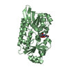
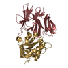
[English] 日本語
Yorodumi
- PDB-6fbr: Crystal Structure of the Human Retinoid X Receptor DNA-Binding Do... -

+
Open data


ID or keywords:

Loading...

-
Basic information

Entry
Database: PDB / ID: 6fbr
TitleCrystal Structure of the Human Retinoid X Receptor DNA-Binding Domain Bound to the Human MEp DR1 Response Element, pH 4.2
Components
  • DNA (5'-D(*CP*TP*GP*GP*GP*TP*CP*AP*AP*AP*GP*TP*TP*CP*AP*TP*C)-3')
  • DNA (5'-D(*GP*AP*TP*GP*AP*AP*CP*TP*TP*TP*GP*AP*CP*CP*CP*AP*G)-3')
  • Retinoic acid receptor RXR-alpha
KeywordsTRANSCRIPTION / TRANSCRIPTION-DNA COMPLEX / NUCLEAR RECEPTOR
Function / homology
Function and homology information


positive regulation of transporter activity / retinoic acid-responsive element binding / NR1H2 & NR1H3 regulate gene expression linked to gluconeogenesis / NR1H2 & NR1H3 regulate gene expression linked to triglyceride lipolysis in adipose / NR1H2 & NR1H3 regulate gene expression to limit cholesterol uptake / positive regulation of thyroid hormone receptor signaling pathway / NR1H2 & NR1H3 regulate gene expression linked to lipogenesis / Carnitine metabolism / ion binding / Regulation of pyruvate dehydrogenase (PDH) complex ...positive regulation of transporter activity / retinoic acid-responsive element binding / NR1H2 & NR1H3 regulate gene expression linked to gluconeogenesis / NR1H2 & NR1H3 regulate gene expression linked to triglyceride lipolysis in adipose / NR1H2 & NR1H3 regulate gene expression to limit cholesterol uptake / positive regulation of thyroid hormone receptor signaling pathway / NR1H2 & NR1H3 regulate gene expression linked to lipogenesis / Carnitine metabolism / ion binding / Regulation of pyruvate dehydrogenase (PDH) complex / retinoic acid binding / positive regulation of vitamin D receptor signaling pathway / nuclear vitamin D receptor binding / Signaling by Retinoic Acid / DNA binding domain binding / nuclear steroid receptor activity / NR1H2 & NR1H3 regulate gene expression to control bile acid homeostasis / LBD domain binding / Synthesis of bile acids and bile salts / positive regulation of cholesterol efflux / retinoic acid receptor signaling pathway / Endogenous sterols / Synthesis of bile acids and bile salts via 27-hydroxycholesterol / positive regulation of bone mineralization / Synthesis of bile acids and bile salts via 7alpha-hydroxycholesterol / response to retinoic acid / Recycling of bile acids and salts / NR1H3 & NR1H2 regulate gene expression linked to cholesterol transport and efflux / peroxisome proliferator activated receptor signaling pathway / RORA activates gene expression / Regulation of lipid metabolism by PPARalpha / hormone-mediated signaling pathway / BMAL1:CLOCK,NPAS2 activates circadian gene expression / Activation of gene expression by SREBF (SREBP) / transcription coregulator binding / nuclear receptor binding / RNA polymerase II transcription regulatory region sequence-specific DNA binding / peptide binding / Heme signaling / SUMOylation of intracellular receptors / mRNA transcription by RNA polymerase II / Transcriptional activation of mitochondrial biogenesis / PPARA activates gene expression / Cytoprotection by HMOX1 / Transcriptional regulation of white adipocyte differentiation / Activation of anterior HOX genes in hindbrain development during early embryogenesis / Nuclear Receptor transcription pathway / Transcriptional regulation of granulopoiesis / RNA polymerase II transcription regulator complex / nuclear receptor activity / sequence-specific double-stranded DNA binding / Circadian Clock / double-stranded DNA binding / transcription regulator complex / sequence-specific DNA binding / cell differentiation / receptor complex / transcription cis-regulatory region binding / DNA-binding transcription factor activity, RNA polymerase II-specific / RNA polymerase II cis-regulatory region sequence-specific DNA binding / DNA-binding transcription factor activity / chromatin / positive regulation of DNA-templated transcription / enzyme binding / negative regulation of transcription by RNA polymerase II / positive regulation of transcription by RNA polymerase II / mitochondrion / zinc ion binding / nucleoplasm / identical protein binding / nucleus / cytosol
Similarity search - Function
Erythroid Transcription Factor GATA-1, subunit A / Erythroid Transcription Factor GATA-1; Chain A / Nuclear/hormone receptor activator site AF-1 / Nuclear/hormone receptor activator site AF-1 / Retinoid X receptor/HNF4 / Nuclear hormone receptor / Nuclear hormones receptors DNA-binding region signature. / Zinc finger, nuclear hormone receptor-type / Zinc finger, C4 type (two domains) / Nuclear hormone receptors DNA-binding domain profile. ...Erythroid Transcription Factor GATA-1, subunit A / Erythroid Transcription Factor GATA-1; Chain A / Nuclear/hormone receptor activator site AF-1 / Nuclear/hormone receptor activator site AF-1 / Retinoid X receptor/HNF4 / Nuclear hormone receptor / Nuclear hormones receptors DNA-binding region signature. / Zinc finger, nuclear hormone receptor-type / Zinc finger, C4 type (two domains) / Nuclear hormone receptors DNA-binding domain profile. / c4 zinc finger in nuclear hormone receptors / Nuclear hormone receptor, ligand-binding domain / Nuclear hormone receptor-like domain superfamily / Ligand-binding domain of nuclear hormone receptor / Nuclear receptor (NR) ligand-binding (LBD) domain profile. / Ligand binding domain of hormone receptors / Zinc finger, NHR/GATA-type / 2-Layer Sandwich / Alpha Beta
Similarity search - Domain/homology
DI(HYDROXYETHYL)ETHER / TRIETHYLENE GLYCOL / PHOSPHATE ION / DNA / DNA (> 10) / Retinoic acid receptor RXR-alpha
Similarity search - Component
Biological speciesHomo sapiens (human)
MethodX-RAY DIFFRACTION / SYNCHROTRON / MOLECULAR REPLACEMENT / molecular replacement / Resolution: 2.1 Å
AuthorsMcEwen, A.G. / Poussin-Courmontagne, P. / Osz, J. / Rochel, N.
Funding support France, 4items
OrganizationGrant numberCountry
French National Research AgencyANR-13-BSV8-0024-01 France
Fondation ARCSFI20121205585 France
French National Research AgencyANR-10-INSB-05-01 France
French National Research AgencyANR-10-LABX-0030-INRT France
CitationJournal: Mol. Cell. Endocrinol. / Year: 2019
Title: Modulation of RXR-DNA complex assembly by DNA context.
Authors: Osz, J. / McEwen, A.G. / Wolf, J. / Poussin-Courmontagne, P. / Peluso-Iltis, C. / Chebaro, Y. / Kieffer, B. / Rochel, N.
History
DepositionDec 19, 2017Deposition site: PDBE / Processing site: PDBE
Revision 1.0Dec 5, 2018Provider: repository / Type: Initial release
Revision 1.1Jan 9, 2019Group: Data collection / Database references
Category: citation / pdbx_database_proc / pdbx_seq_map_depositor_info
Item: _citation.journal_volume / _citation.page_first ..._citation.journal_volume / _citation.page_first / _citation.page_last / _citation.year / _pdbx_seq_map_depositor_info.one_letter_code_mod
Revision 1.2Jan 17, 2024Group: Data collection / Database references / Refinement description
Category: chem_comp_atom / chem_comp_bond ...chem_comp_atom / chem_comp_bond / database_2 / pdbx_initial_refinement_model
Item: _database_2.pdbx_DOI / _database_2.pdbx_database_accession

-
Structure visualization

Structure viewerMolecule:
MolmilJmol/JSmol

Downloads & links

-
Assembly

Deposited unit
A: Retinoic acid receptor RXR-alpha
B: Retinoic acid receptor RXR-alpha
C: DNA (5'-D(*CP*TP*GP*GP*GP*TP*CP*AP*AP*AP*GP*TP*TP*CP*AP*TP*C)-3')
D: DNA (5'-D(*GP*AP*TP*GP*AP*AP*CP*TP*TP*TP*GP*AP*CP*CP*CP*AP*G)-3')
hetero molecules


Theoretical massNumber of molelcules
Total (without water)31,52810
Polymers30,9404
Non-polymers5886
Water2,414134
1


  • Idetical with deposited unit
  • defined by author&software
  • Evidence: gel filtration, mass spectrometry, isothermal titration calorimetry
TypeNameSymmetry operationNumber
identity operation1_555x,y,z1
Buried area6600 Å2
ΔGint-35 kcal/mol
Surface area13350 Å2
MethodPISA
Unit cell
Length a, b, c (Å)55.659, 55.659, 166.131
Angle α, β, γ (deg.)90.000, 90.000, 120.000
Int Tables number152
Space group name H-MP3121
Components on special symmetry positions
IDModelComponents
11B-304-

PEG

-
Components

-
Protein , 1 types, 2 molecules AB

#1: Protein Retinoic acid receptor RXR-alpha / Nuclear receptor subfamily 2 group B member 1 / Retinoid X receptor alpha


Mass: 10262.926 Da / Num. of mol.: 2
Source method: isolated from a genetically manipulated source
Source: (gene. exp.) Homo sapiens (human) / Gene: RXRA, NR2B1 / Production host: Escherichia coli BL21(DE3) (bacteria) / References: UniProt: P19793

-
DNA chain , 2 types, 2 molecules CD

#2: DNA chain DNA (5'-D(*CP*TP*GP*GP*GP*TP*CP*AP*AP*AP*GP*TP*TP*CP*AP*TP*C)-3')


Mass: 5202.383 Da / Num. of mol.: 1 / Source method: obtained synthetically / Source: (synth.) Homo sapiens (human)
#3: DNA chain DNA (5'-D(*GP*AP*TP*GP*AP*AP*CP*TP*TP*TP*GP*AP*CP*CP*CP*AP*G)-3')


Mass: 5211.398 Da / Num. of mol.: 1 / Source method: obtained synthetically / Source: (synth.) Homo sapiens (human)

-
Non-polymers , 5 types, 140 molecules

#4: Chemical ChemComp-ZN / ZINC ION


Mass: 65.409 Da / Num. of mol.: 2 / Source method: obtained synthetically / Formula: Zn
#5: Chemical ChemComp-PO4 / PHOSPHATE ION


Mass: 94.971 Da / Num. of mol.: 1 / Source method: obtained synthetically / Formula: PO4
#6: Chemical ChemComp-PGE / TRIETHYLENE GLYCOL


Mass: 150.173 Da / Num. of mol.: 1 / Source method: obtained synthetically / Formula: C6H14O4
#7: Chemical ChemComp-PEG / DI(HYDROXYETHYL)ETHER


Mass: 106.120 Da / Num. of mol.: 2 / Source method: obtained synthetically / Formula: C4H10O3
#8: Water ChemComp-HOH / water


Mass: 18.015 Da / Num. of mol.: 134 / Source method: isolated from a natural source / Formula: H2O

-
Experimental details

-
Experiment

ExperimentMethod: X-RAY DIFFRACTION / Number of used crystals: 1

-
Sample preparation

CrystalDensity Matthews: 2.4 Å3/Da / Density % sol: 48.77 %
Crystal growTemperature: 293 K / Method: vapor diffusion, sitting drop / pH: 4.2 / Details: 40% PEG 300, 0.1M sodium phosphate citrate pH 4.2

-
Data collection

DiffractionMean temperature: 100 K
Diffraction sourceSource: SYNCHROTRON / Site: ESRF / Beamline: ID30B / Wavelength: 1.2821 Å
DetectorType: DECTRIS PILATUS3 6M / Detector: PIXEL / Date: Jun 13, 2016
RadiationMonochromator: Si(111) / Protocol: SINGLE WAVELENGTH / Monochromatic (M) / Laue (L): M / Scattering type: x-ray
Radiation wavelengthWavelength: 1.2821 Å / Relative weight: 1
ReflectionResolution: 2→46.29 Å / Num. obs: 19015 / % possible obs: 90.2 % / Redundancy: 4.624 % / Biso Wilson estimate: 52.94 Å2 / CC1/2: 0.998 / Rmerge(I) obs: 0.113 / Rrim(I) all: 0.127 / Χ2: 1.224 / Net I/σ(I): 8.79 / Num. measured all: 87918
Reflection shell

Diffraction-ID: 1

Resolution (Å)Redundancy (%)Rmerge(I) obsMean I/σ(I) obsNum. unique obsCC1/2Rrim(I) all% possible all
2-2.124.183.280.3919480.1523.73258.9
2.12-2.274.7041.930.827970.3782.16889
2.27-2.454.7351.0811.5528570.6421.21497.6
2.45-2.684.6790.6392.5126400.8530.71996.7
2.68-34.6760.3224.8424150.9620.36297.9
3-3.464.8910.09312.0921490.9980.10597.6
3.46-4.234.7060.05422.4518410.9990.0698
4.23-5.964.5030.04329.3514720.9980.04998.1
5.96-46.294.0640.03434.418960.9990.03996.3

-
Phasing

PhasingMethod: molecular replacement

-
Processing

Software
NameVersionClassificationNB
XDSdata reduction
XSCALEdata scaling
PHASERphasing
BUSTER2.10.2refinement
PDB_EXTRACT3.24data extraction
RefinementMethod to determine structure: MOLECULAR REPLACEMENT
Starting model: 6FBQ
Resolution: 2.1→46.29 Å / Cor.coef. Fo:Fc: 0.953 / Cor.coef. Fo:Fc free: 0.933 / Rfactor Rfree error: 0 / SU R Cruickshank DPI: 0.187 / Cross valid method: THROUGHOUT / σ(F): 0 / SU R Blow DPI: 0.212 / SU Rfree Blow DPI: 0.182 / SU Rfree Cruickshank DPI: 0.172
RfactorNum. reflection% reflectionSelection details
Rfree0.232 869 5 %RANDOM
Rwork0.178 ---
obs0.181 17378 95.5 %-
Displacement parametersBiso max: 158.57 Å2 / Biso mean: 58.52 Å2 / Biso min: 32.71 Å2
Baniso -1Baniso -2Baniso -3
1--5.7438 Å20 Å20 Å2
2---5.7438 Å20 Å2
3---11.4877 Å2
Refine analyzeLuzzati coordinate error obs: 0.3 Å
Refinement stepCycle: final / Resolution: 2.1→46.29 Å
ProteinNucleic acidLigandSolventTotal
Num. atoms1284 691 31 134 2140
Biso mean--76.28 57.03 -
Num. residues----193
Refine LS restraints
Refine-IDTypeNumberRestraint functionWeightDev ideal
X-RAY DIFFRACTIONt_dihedral_angle_d698SINUSOIDAL2
X-RAY DIFFRACTIONt_trig_c_planes27HARMONIC2
X-RAY DIFFRACTIONt_gen_planes235HARMONIC5
X-RAY DIFFRACTIONt_it2128HARMONIC20
X-RAY DIFFRACTIONt_nbd0SEMIHARMONIC5
X-RAY DIFFRACTIONt_improper_torsion
X-RAY DIFFRACTIONt_pseud_angle
X-RAY DIFFRACTIONt_chiral_improper_torsion265SEMIHARMONIC5
X-RAY DIFFRACTIONt_sum_occupancies
X-RAY DIFFRACTIONt_utility_distance
X-RAY DIFFRACTIONt_utility_angle
X-RAY DIFFRACTIONt_utility_torsion
X-RAY DIFFRACTIONt_ideal_dist_contact2261SEMIHARMONIC4
X-RAY DIFFRACTIONt_bond_d2128HARMONIC20.01
X-RAY DIFFRACTIONt_angle_deg2977HARMONIC21.06
X-RAY DIFFRACTIONt_omega_torsion2.9
X-RAY DIFFRACTIONt_other_torsion20.49
LS refinement shellResolution: 2.1→2.23 Å / Rfactor Rfree error: 0 / Total num. of bins used: 9
RfactorNum. reflection% reflection
Rfree0.268 121 5 %
Rwork0.235 2297 -
all0.236 2418 -
obs--83.71 %
Refinement TLS params.

Method: refined / Refine-ID: X-RAY DIFFRACTION

IDL112)L122)L132)L222)L232)L332)S11 (Å °)S12 (Å °)S13 (Å °)S21 (Å °)S22 (Å °)S23 (Å °)S31 (Å °)S32 (Å °)S33 (Å °)T112)T122)T132)T222)T232)T332)Origin x (Å)Origin y (Å)Origin z (Å)
15.1928-1.14210.28232.6856-0.51335.6509-0.0733-0.12990.2834-0.0120.12640.0826-0.4429-0.072-0.0531-0.0873-0.0427-0.0273-0.07130.0486-0.1494-23.189924.54219.9389
22.18981.0478-0.18234.8217-1.684.77610.0111-0.0186-0.19910.3368-0.0409-0.25180.0660.34770.0297-0.05010.0057-0.0081-0.0516-0.0079-0.1726-19.55346.615935.6569
30.78890.35650.94350.8066-2.35442.77430.04480.08580.0792-0.16930.16290.00050.118-0.3866-0.20770.0154-0.05630.00130.0446-0.0301-0.2027-29.583913.650721.0803
40.7337-0.16390.64381.0818-1.184.59510.07150.01690.04090.02760.03890.03480.337-0.2017-0.1104-0.0178-0.0524-0.01250.0351-0.0248-0.1736-28.477911.915421.635
Refinement TLS group
IDRefine-IDRefine TLS-IDSelection detailsAuth asym-IDAuth seq-ID
1X-RAY DIFFRACTION1{ A|* }A131 - 208
2X-RAY DIFFRACTION2{ B|* }B126 - 210
3X-RAY DIFFRACTION3{ C|* }C1 - 17
4X-RAY DIFFRACTION4{ D|* }D1 - 17

+
About Yorodumi

-
News

-
Feb 9, 2022. New format data for meta-information of EMDB entries

New format data for meta-information of EMDB entries

  • Version 3 of the EMDB header file is now the official format.
  • The previous official version 1.9 will be removed from the archive.

Related info.:EMDB header

External links:wwPDB to switch to version 3 of the EMDB data model

-
Aug 12, 2020. Covid-19 info

Covid-19 info

URL: https://pdbjlvh1.pdbj.org/emnavi/covid19.php

New page: Covid-19 featured information page in EM Navigator.

Related info.:Covid-19 info / Mar 5, 2020. Novel coronavirus structure data

+
Mar 5, 2020. Novel coronavirus structure data

Novel coronavirus structure data

Related info.:Yorodumi Speices / Aug 12, 2020. Covid-19 info

External links:COVID-19 featured content - PDBj / Molecule of the Month (242):Coronavirus Proteases

+
Jan 31, 2019. EMDB accession codes are about to change! (news from PDBe EMDB page)

EMDB accession codes are about to change! (news from PDBe EMDB page)

  • The allocation of 4 digits for EMDB accession codes will soon come to an end. Whilst these codes will remain in use, new EMDB accession codes will include an additional digit and will expand incrementally as the available range of codes is exhausted. The current 4-digit format prefixed with “EMD-” (i.e. EMD-XXXX) will advance to a 5-digit format (i.e. EMD-XXXXX), and so on. It is currently estimated that the 4-digit codes will be depleted around Spring 2019, at which point the 5-digit format will come into force.
  • The EM Navigator/Yorodumi systems omit the EMD- prefix.

Related info.:Q: What is EMD? / ID/Accession-code notation in Yorodumi/EM Navigator

External links:EMDB Accession Codes are Changing Soon! / Contact to PDBj

+
Jul 12, 2017. Major update of PDB

Major update of PDB

  • wwPDB released updated PDB data conforming to the new PDBx/mmCIF dictionary.
  • This is a major update changing the version number from 4 to 5, and with Remediation, in which all the entries are updated.
  • In this update, many items about electron microscopy experimental information are reorganized (e.g. em_software).
  • Now, EM Navigator and Yorodumi are based on the updated data.

External links:wwPDB Remediation / Enriched Model Files Conforming to OneDep Data Standards Now Available in the PDB FTP Archive

-
Yorodumi

Thousand views of thousand structures

  • Yorodumi is a browser for structure data from EMDB, PDB, SASBDB, etc.
  • This page is also the successor to EM Navigator detail page, and also detail information page/front-end page for Omokage search.
  • The word "yorodu" (or yorozu) is an old Japanese word meaning "ten thousand". "mi" (miru) is to see.

Related info.:EMDB / PDB / SASBDB / Comparison of 3 databanks / Yorodumi Search / Aug 31, 2016. New EM Navigator & Yorodumi / Yorodumi Papers / Jmol/JSmol / Function and homology information / Changes in new EM Navigator and Yorodumi

Read more