PROTON TRANSPORT / electron cryo-microscopy mitochondrial ATP synthase membrane protein energy conversion proton pathway
Function / homology
Function and homology information
proton transmembrane transporter activity / proton motive force-driven ATP synthesis / proton-transporting two-sector ATPase complex, proton-transporting domain / proton-transporting ATP synthase complex / proton-transporting ATP synthase activity, rotational mechanism / lipid binding / mitochondrion Similarity search - Function
F1F0 ATP synthase subunit C / F1FO ATP Synthase / ATP synthase, F0 complex, subunit A, bacterial/mitochondria / ATP synthase, F0 complex, subunit A / ATP synthase, F0 complex, subunit A, active site / ATP synthase, F0 complex, subunit A superfamily / ATP synthase A chain / ATP synthase a subunit signature. / ATP synthase, F0 complex, subunit C / F1F0 ATP synthase subunit C superfamily ...F1F0 ATP synthase subunit C / F1FO ATP Synthase / ATP synthase, F0 complex, subunit A, bacterial/mitochondria / ATP synthase, F0 complex, subunit A / ATP synthase, F0 complex, subunit A, active site / ATP synthase, F0 complex, subunit A superfamily / ATP synthase A chain / ATP synthase a subunit signature. / ATP synthase, F0 complex, subunit C / F1F0 ATP synthase subunit C superfamily / ATP synthase, F0 complex, subunit C, DCCD-binding site / ATP synthase c subunit signature. / V-ATPase proteolipid subunit C-like domain / F/V-ATP synthase subunit C superfamily / ATP synthase subunit C / Up-down Bundle / Mainly Alpha Similarity search - Domain/homology
Mitochondrial ATP synthase subunit c / Mitochondrial ATP synthase subunit ASA6 / F-ATPase protein 6 Similarity search - Component
Biological species
Polytomella sp. Pringsheim 198.80 (plant)
Method
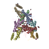
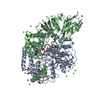
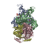
ELECTRON MICROSCOPY / single particle reconstruction / cryo EM / Resolution: 3.7 Å
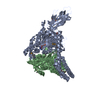
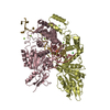
Journal: Elife / Year: 2017 Title: Structural basis of proton translocation and force generation in mitochondrial ATP synthase. Authors: Niklas Klusch / Bonnie J Murphy / Deryck J Mills / Özkan Yildiz / Werner Kühlbrandt / Abstract: ATP synthases produce ATP by rotary catalysis, powered by the electrochemical proton gradient across the membrane. Understanding this fundamental process requires an atomic model of the proton ...ATP synthases produce ATP by rotary catalysis, powered by the electrochemical proton gradient across the membrane. Understanding this fundamental process requires an atomic model of the proton pathway. We determined the structure of an intact mitochondrial ATP synthase dimer by electron cryo-microscopy at near-atomic resolution. Charged and polar residues of the -subunit stator define two aqueous channels, each spanning one half of the membrane. Passing through a conserved membrane-intrinsic helix hairpin, the lumenal channel protonates an acidic glutamate in the -ring rotor. Upon ring rotation, the protonated glutamate encounters the matrix channel and deprotonates. An arginine between the two channels prevents proton leakage. The steep potential gradient over the sub-nm inter-channel distance exerts a force on the deprotonated glutamate, resulting in net directional rotation.
History
Deposition
Nov 28, 2017
Deposition site: PDBE / Processing site: PDBE
Revision 1.0
Dec 20, 2017
Provider: repository / Type: Initial release
Revision 1.0
Dec 20, 2017
Data content type: EM metadata / Data content type: EM metadata / Provider: repository / Type: Initial release
Revision 1.0
Dec 20, 2017
Data content type: Image / Data content type: Image / Provider: repository / Type: Initial release
Revision 1.0
Dec 20, 2017
Data content type: Primary map / Data content type: Primary map / Provider: repository / Type: Initial release
Revision 1.0
Dec 20, 2017
Data content type: Image / Data content type: Image / Provider: repository / Type: Initial release
Revision 1.0
Dec 20, 2017
Data content type: Primary map / Data content type: Primary map / Provider: repository / Type: Initial release
Revision 1.0
Dec 20, 2017
Data content type: Image / Data content type: Image / Provider: repository / Type: Initial release
Revision 1.0
Dec 20, 2017
Data content type: Primary map / Data content type: Primary map / Provider: repository / Type: Initial release
Revision 1.0
Dec 20, 2017
Data content type: Image / Data content type: Image / Provider: repository / Type: Initial release
Revision 1.0
Dec 20, 2017
Data content type: Primary map / Data content type: Primary map / Provider: repository / Type: Initial release
Data content type: EM metadata / Data content type: EM metadata / EM metadata / Group: Data processing / Experimental summary / Data content type: EM metadata / EM metadata / Category: em_admin / em_software / Data content type: EM metadata / EM metadata / Item: _em_admin.last_update / _em_software.name
A: Mitochondrial ATP synthase subunit c B: Mitochondrial ATP synthase subunit c C: Mitochondrial ATP synthase subunit c D: Mitochondrial ATP synthase subunit c E: Mitochondrial ATP synthase subunit c F: Mitochondrial ATP synthase subunit c G: Mitochondrial ATP synthase subunit c H: Mitochondrial ATP synthase subunit c I: Mitochondrial ATP synthase subunit c J: Mitochondrial ATP synthase subunit c M: Mitochondrial ATP synthase subunit 6 N: Mitochondrial ATP synthase subunit ASA6
Instrument: FEI VITROBOT MARK II / Cryogen name: ETHANE / Humidity: 70 % / Chamber temperature: 282 K
-
Electron microscopy imaging
Microscopy
Model: JEOL 3200FSC
Electron gun
Electron source: FIELD EMISSION GUN / Accelerating voltage: 300 kV / Illumination mode: FLOOD BEAM
Electron lens
Mode: BRIGHT FIELD
Image recording
Average exposure time: 0.2 sec. / Electron dose: 82 e/Å2 / Detector mode: COUNTING / Film or detector model: GATAN K2 SUMMIT (4k x 4k) / Num. of grids imaged: 30
Image scans
Width: 4096 / Height: 4096 / Movie frames/image: 45 / Used frames/image: 1-45
In the structure databanks used in Yorodumi, some data are registered as the other names, "COVID-19 virus" and "2019-nCoV". Here are the details of the virus and the list of structure data.
Jan 31, 2019. EMDB accession codes are about to change! (news from PDBe EMDB page)
EMDB accession codes are about to change! (news from PDBe EMDB page)
The allocation of 4 digits for EMDB accession codes will soon come to an end. Whilst these codes will remain in use, new EMDB accession codes will include an additional digit and will expand incrementally as the available range of codes is exhausted. The current 4-digit format prefixed with “EMD-” (i.e. EMD-XXXX) will advance to a 5-digit format (i.e. EMD-XXXXX), and so on. It is currently estimated that the 4-digit codes will be depleted around Spring 2019, at which point the 5-digit format will come into force.
The EM Navigator/Yorodumi systems omit the EMD- prefix.
Related info.:Q: What is EMD? / ID/Accession-code notation in Yorodumi/EM Navigator
Yorodumi is a browser for structure data from EMDB, PDB, SASBDB, etc.
This page is also the successor to EM Navigator detail page, and also detail information page/front-end page for Omokage search.
The word "yorodu" (or yorozu) is an old Japanese word meaning "ten thousand". "mi" (miru) is to see.
Related info.:EMDB / PDB / SASBDB / Comparison of 3 databanks / Yorodumi Search / Aug 31, 2016. New EM Navigator & Yorodumi / Yorodumi Papers / Jmol/JSmol / Function and homology information / Changes in new EM Navigator and Yorodumi
 Movie
Movie Controller


 Open data
 Basic information
 Components
 Keywords
 Function and homology information
 Polytomella sp. Pringsheim 198.80 (plant)
 Authors
 Germany, 3items
  Citation
 Structure visualization
 Downloads & links
 Other downloads
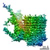


 PDBj
PDBj

 Assembly
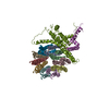
 Sample preparation
 Electron microscopy imaging
 FIELD EMISSION GUN / Accelerating voltage: 300 kV / Illumination mode: FLOOD BEAM
 Processing