 Movie
Movie Controller

[English] 日本語
 Yorodumi
- PDB-6eyv: Crystal structure of the pyoverdine maturation protein PvdP in co... -
+ Open data

- Basic information
| Entry | Database: PDB / ID: 6eyv | ||||||
|---|---|---|---|---|---|---|---|
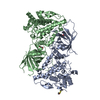
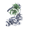
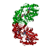
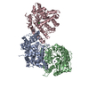
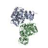
| Title | Crystal structure of the pyoverdine maturation protein PvdP in complex with the mock substrates L-tyrosine and zinc. | ||||||
 Components Components | PvdP | ||||||
 Keywords Keywords | OXIDOREDUCTASE / tyrosinase / pyoverdine / barrel / zinc | ||||||
| Function / homology | Di-copper centre-containing domain superfamily / Twin-arginine translocation pathway, signal sequence, bacterial/archaeal / metal ion binding / TYROSINE / PvdP Function and homology information Function and homology information | ||||||
| Biological species |   Pseudomonas aeruginosa (bacteria) Pseudomonas aeruginosa (bacteria) | ||||||
| Method |  X-RAY DIFFRACTION / SYNCHROTRON / MOLECULAR REPLACEMENT / Resolution: 2.704 Å X-RAY DIFFRACTION / SYNCHROTRON / MOLECULAR REPLACEMENT / Resolution: 2.704 Å | ||||||
 Authors Authors | Poppe, J. / Blankenfeldt, W. | ||||||
 Citation Citation | Journal: J. Biol. Chem. / Year: 2018 Title: Pseudomonas aeruginosapyoverdine maturation enzyme PvdP has a noncanonical domain architecture and affords insight into a new subclass of tyrosinases. Authors: Poppe, J. / Reichelt, J. / Blankenfeldt, W. | ||||||
| History |
|
- Structure visualization
| Structure viewer | Molecule: MolmilJmol/JSmol |
|---|
- Downloads & links
-Download
| PDBx/mmCIF format | 6eyv.cif.gz | 343.4 KB | Display | PDBx/mmCIF format |
|---|---|---|---|---|
| PDB format | pdb6eyv.ent.gz | 279.7 KB | Display | PDB format |
| PDBx/mmJSON format | 6eyv.json.gz | Tree view | PDBx/mmJSON format | |
| Others |  Other downloads Other downloads |
-Validation report
| Summary document | 6eyv_validation.pdf.gz | 450.3 KB | Display | wwPDB validaton report |
|---|---|---|---|---|
| Full document | 6eyv_full_validation.pdf.gz | 451.3 KB | Display | |
| Data in XML | 6eyv_validation.xml.gz | 30.9 KB | Display | |
| Data in CIF | 6eyv_validation.cif.gz | 41.6 KB | Display | |
| Arichive directory | https://data.pdbj.org/pub/pdb/validation_reports/ey/6eyvftp://data.pdbj.org/pub/pdb/validation_reports/ey/6eyv | HTTPS FTP |
-Related structure data
| Related structure data |  6eysSC S: Starting model for refinement C: citing same article ( |
|---|---|
| Similar structure data |

-Links
 PDBj
PDBj


- Assembly
| Deposited unit | 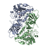
| ||||||||
|---|---|---|---|---|---|---|---|---|---|
| 1 |
| ||||||||
| Unit cell |
|
-Components
| #1: Protein | Mass: 61758.133 Da / Num. of mol.: 2 Source method: isolated from a genetically manipulated source Source: (gene. exp.) Pseudomonas aeruginosa (strain UCBPP-PA14) (bacteria)Strain: UCBPP-PA14 / Gene: pvdP, PA14_33740 / Production host: #2: Chemical | ChemComp-ZN /   Mass: 65.409 Da / Num. of mol.: 4 / Source method: obtained synthetically / Formula: Zn Mass: 65.409 Da / Num. of mol.: 4 / Source method: obtained synthetically / Formula: Zn#3: Chemical |   Type: L-peptide linking / Mass: 181.189 Da / Num. of mol.: 2 / Source method: obtained synthetically / Formula: C9H11NO3 Type: L-peptide linking / Mass: 181.189 Da / Num. of mol.: 2 / Source method: obtained synthetically / Formula: C9H11NO3#4: Water | ChemComp-HOH / |  Mass: 18.015 Da / Num. of mol.: 24 / Source method: isolated from a natural source / Formula: H2O Mass: 18.015 Da / Num. of mol.: 24 / Source method: isolated from a natural source / Formula: H2O |
|---|
-Experimental details
-Experiment
| Experiment | Method: X-RAY DIFFRACTION / Number of used crystals: 1 |
|---|
- Sample preparation
| Crystal | Density Matthews: 2.81 Å3/Da / Density % sol: 56.16 % / Description: thin plates |
|---|---|
| Crystal grow | Temperature: 293.15 K / Method: vapor diffusion, sitting drop Details: 0.857 M NH4SO4 0.1 M MES pH 5.5 0.5 M ZnCl2 1 mM L-tyrosine |
-Data collection
| Diffraction | Mean temperature: 100 K |
|---|---|
| Diffraction source | Source: SYNCHROTRON / Site: BESSY  / Beamline: 14.2 / Wavelength: 1.282 Å / Beamline: 14.2 / Wavelength: 1.282 Å |
| Detector | Type: DECTRIS PILATUS3 2M / Detector: PIXEL / Date: Jan 13, 2017 |
| Radiation | Protocol: SINGLE WAVELENGTH / Monochromatic (M) / Laue (L): M / Scattering type: x-ray |
| Radiation wavelength | Wavelength: 1.282 Å / Relative weight: 1 |
| Reflection | Resolution: 2.701→48.092 Å / Num. obs: 23084 / % possible obs: 92.2 % / Redundancy: 9.9 % / CC1/2: 0.988 / Rpim(I) all: 0.101 / Rrim(I) all: 0.32 / Net I/σ(I): 8.6 |
| Reflection shell | Resolution: 2.701→3.032 Å / Redundancy: 7.5 % / Mean I/σ(I) obs: 1.6 / Num. measured obs: 8710 / Num. unique obs: 1154 / CC1/2: 0.506 / Rpim(I) all: 0.536 / % possible all: 56.5 |
- Processing
| Software |
| |||||||||||||||||||||||||||||||||||||||||||||||||||||||||||||||||||||||||||||||||||||||||||||||||||||||||
|---|---|---|---|---|---|---|---|---|---|---|---|---|---|---|---|---|---|---|---|---|---|---|---|---|---|---|---|---|---|---|---|---|---|---|---|---|---|---|---|---|---|---|---|---|---|---|---|---|---|---|---|---|---|---|---|---|---|---|---|---|---|---|---|---|---|---|---|---|---|---|---|---|---|---|---|---|---|---|---|---|---|---|---|---|---|---|---|---|---|---|---|---|---|---|---|---|---|---|---|---|---|---|---|---|---|---|
| Refinement | Method to determine structure: MOLECULAR REPLACEMENT Starting model: 6EYS Resolution: 2.704→48.092 Å / SU ML: 0.32 / Cross valid method: FREE R-VALUE / σ(F): 1.34 / Phase error: 27.55
| |||||||||||||||||||||||||||||||||||||||||||||||||||||||||||||||||||||||||||||||||||||||||||||||||||||||||
| Solvent computation | Shrinkage radii: 0.9 Å / VDW probe radii: 1.11 Å | |||||||||||||||||||||||||||||||||||||||||||||||||||||||||||||||||||||||||||||||||||||||||||||||||||||||||
| Refinement step | Cycle: LAST / Resolution: 2.704→48.092 Å
| |||||||||||||||||||||||||||||||||||||||||||||||||||||||||||||||||||||||||||||||||||||||||||||||||||||||||
| Refine LS restraints |
| |||||||||||||||||||||||||||||||||||||||||||||||||||||||||||||||||||||||||||||||||||||||||||||||||||||||||
| LS refinement shell |
|
