 Movie
Movie Controller

[English] 日本語
 Yorodumi
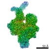
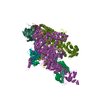
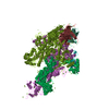
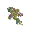
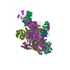
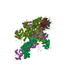
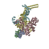
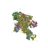
- PDB-6eyd: Structure of Mycobacterium smegmatis RNA polymerase Sigma-A holoenzyme -
+ Open data

- Basic information
| Entry | Database: PDB / ID: 6eyd | |||||||||
|---|---|---|---|---|---|---|---|---|---|---|
| Title | Structure of Mycobacterium smegmatis RNA polymerase Sigma-A holoenzyme | |||||||||
 Components Components |
| |||||||||
 Keywords Keywords | TRANSCRIPTION / TRANSCRIPTION Sigma | |||||||||
| Function / homology |  Function and homology information Function and homology informationsigma factor activity / DNA-directed RNA polymerase complex / DNA-templated transcription initiation / ribonucleoside binding / DNA-directed RNA polymerase / DNA-directed RNA polymerase activity / protein dimerization activity / response to antibiotic / magnesium ion binding / DNA-templated transcription ...sigma factor activity / DNA-directed RNA polymerase complex / DNA-templated transcription initiation / ribonucleoside binding / DNA-directed RNA polymerase / DNA-directed RNA polymerase activity / protein dimerization activity / response to antibiotic / magnesium ion binding / DNA-templated transcription / DNA binding / zinc ion binding / cytoplasm Similarity search - Function | |||||||||
| Biological species |  Mycobacterium smegmatis (bacteria) Mycobacterium smegmatis (bacteria) | |||||||||
| Method | ELECTRON MICROSCOPY / single particle reconstruction / cryo EM / Resolution: 4.22 Å | |||||||||
 Authors Authors | Kouba, T. / Barvik, I. / Krasny, L. | |||||||||
 Citation Citation | Journal: J Bacteriol / Year: 2019 Title: The Core and Holoenzyme Forms of RNA Polymerase from . Authors: Tomáš Kouba / Jiří Pospíšil / Jarmila Hnilicová / Hana Šanderová / Ivan Barvík / Libor Krásný /   Abstract: Bacterial RNA polymerase (RNAP) is essential for gene expression and as such is a valid drug target. Hence, it is imperative to know its structure and dynamics. Here, we present two as-yet-unreported ...Bacterial RNA polymerase (RNAP) is essential for gene expression and as such is a valid drug target. Hence, it is imperative to know its structure and dynamics. Here, we present two as-yet-unreported forms of RNAP: core and holoenzyme containing σ but no other factors. Each form was detected by cryo-electron microscopy in two major conformations. Comparisons of these structures with known structures of other RNAPs reveal a high degree of conformational flexibility of the mycobacterial enzyme and confirm that region 1.1 of σ is directed into the primary channel of RNAP. Taken together, we describe the conformational changes of unrestrained mycobacterial RNAP. We describe here three-dimensional structures of core and holoenzyme forms of mycobacterial RNA polymerase (RNAP) solved by cryo-electron microscopy. These structures fill the thus-far-empty spots in the gallery of the pivotal forms of mycobacterial RNAP and illuminate the extent of conformational dynamics of this enzyme. The presented findings may facilitate future designs of antimycobacterial drugs targeting RNAP. | |||||||||
| History |
|
- Structure visualization
| Movie |
Movie viewer |
|---|---|
| Structure viewer | Molecule: MolmilJmol/JSmol |
- Downloads & links
-Download
| PDBx/mmCIF format | 6eyd.cif.gz | 594.2 KB | Display | PDBx/mmCIF format |
|---|---|---|---|---|
| PDB format | pdb6eyd.ent.gz | 462.3 KB | Display | PDB format |
| PDBx/mmJSON format | 6eyd.json.gz | Tree view | PDBx/mmJSON format | |
| Others |  Other downloads Other downloads |
-Validation report
| Arichive directory | https://data.pdbj.org/pub/pdb/validation_reports/ey/6eydftp://data.pdbj.org/pub/pdb/validation_reports/ey/6eyd | HTTPS FTP |
|---|
-Related structure data
| Related structure data | 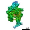 3983MC 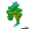 4192C 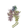 6f6wC M: map data used to model this data C: citing same article ( |
|---|---|
| Similar structure data |
-Links
 PDBj
PDBj

- Assembly
| Deposited unit | 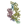
|
|---|---|
| 1 |
|
-Components
-DNA-directed RNA polymerase subunit ... , 4 types, 5 molecules ABCDE
| #1: Protein | Mass: 37959.441 Da / Num. of mol.: 2 Source method: isolated from a genetically manipulated source Source: (gene. exp.) Mycobacterium smegmatis (strain ATCC 700084 / mc(2)155) (bacteria)Gene: rpoA, MSMEG_1524, MSMEI_1488 / Production host: #2: Protein | | Mass: 129456.938 Da / Num. of mol.: 1 Source method: isolated from a genetically manipulated source Source: (gene. exp.) Mycobacterium smegmatis (strain ATCC 700084 / mc(2)155) (bacteria)Gene: rpoB, MSMEG_1367, MSMEI_1328 / Production host: #3: Protein | | Mass: 147785.953 Da / Num. of mol.: 1 Source method: isolated from a genetically manipulated source Source: (gene. exp.) Mycobacterium smegmatis (strain ATCC 700084 / mc(2)155) (bacteria)Gene: rpoC, MSMEG_1368, MSMEI_1329 / Production host: #4: Protein | | Mass: 11512.698 Da / Num. of mol.: 1 Source method: isolated from a genetically manipulated source Source: (gene. exp.) Mycobacterium smegmatis (strain ATCC 700084 / mc(2)155) (bacteria)Gene: rpoZ, MSMEG_3053, MSMEI_2977 / Production host: |
|---|
-Protein , 1 types, 1 molecules F
| #5: Protein | Mass: 51573.551 Da / Num. of mol.: 1 Source method: isolated from a genetically manipulated source Source: (gene. exp.) Mycobacterium smegmatis (strain ATCC 700084 / mc(2)155) (bacteria)Gene: sigA, MSMEG_2758 / Production host: |
|---|
-Non-polymers , 2 types, 3 molecules 

| #6: Chemical |  Mass: 65.409 Da / Num. of mol.: 2 / Source method: obtained synthetically / Formula: Zn Mass: 65.409 Da / Num. of mol.: 2 / Source method: obtained synthetically / Formula: Zn#7: Chemical | ChemComp-MG / |  Mass: 24.305 Da / Num. of mol.: 1 / Source method: obtained synthetically / Formula: Mg Mass: 24.305 Da / Num. of mol.: 1 / Source method: obtained synthetically / Formula: Mg |
|---|
-Experimental details
-Experiment
| Experiment | Method: ELECTRON MICROSCOPY |
|---|---|
| EM experiment | Aggregation state: PARTICLE / 3D reconstruction method: single particle reconstruction |
- Sample preparation
| Component | Name: Mycobacterium smegmatis RNA polymerase Sigma-A holoenzyme Type: COMPLEX / Entity ID: #1-#5 / Source: RECOMBINANT |
|---|---|
| Molecular weight | Value: 0.415 MDa / Experimental value: NO |
| Source (natural) | Organism: Mycobacterium smegmatis str. MC2 155 (bacteria) |
| Source (recombinant) | Organism: |
| Buffer solution | pH: 7.8 |
| Specimen | Embedding applied: NO / Shadowing applied: NO / Staining applied: NO / Vitrification applied: YES |
| Specimen support | Grid material: GRAPHENE OXIDE |
| Vitrification | Cryogen name: ETHANE |
- Electron microscopy imaging
| Experimental equipment |  Model: Tecnai Polara / Image courtesy: FEI Company |
|---|---|
| Microscopy | Model: FEI POLARA 300 |
| Electron gun | Electron source:  FIELD EMISSION GUN / Accelerating voltage: 300 kV / Illumination mode: FLOOD BEAM FIELD EMISSION GUN / Accelerating voltage: 300 kV / Illumination mode: FLOOD BEAM |
| Electron lens | Mode: BRIGHT FIELD / Nominal magnification: 78000 X / Nominal defocus max: 3800 nm / Nominal defocus min: 1500 nm / Cs: 2 mm / C2 aperture diameter: 50 µm / Alignment procedure: COMA FREE |
| Image recording | Average exposure time: 2 sec. / Electron dose: 52 e/Å2 / Detector mode: INTEGRATING / Film or detector model: FEI FALCON III (4k x 4k) |
- Processing
| Software | Name: REFMAC / Version: 5.8.0238 / Classification: refinement | ||||||||||||||||||||||||||||||||||||||||||||||||||||||||||||||||||||||||||||||||||||||||||||||||||||||||||
|---|---|---|---|---|---|---|---|---|---|---|---|---|---|---|---|---|---|---|---|---|---|---|---|---|---|---|---|---|---|---|---|---|---|---|---|---|---|---|---|---|---|---|---|---|---|---|---|---|---|---|---|---|---|---|---|---|---|---|---|---|---|---|---|---|---|---|---|---|---|---|---|---|---|---|---|---|---|---|---|---|---|---|---|---|---|---|---|---|---|---|---|---|---|---|---|---|---|---|---|---|---|---|---|---|---|---|---|
| EM software |
| ||||||||||||||||||||||||||||||||||||||||||||||||||||||||||||||||||||||||||||||||||||||||||||||||||||||||||
| CTF correction | Type: PHASE FLIPPING ONLY | ||||||||||||||||||||||||||||||||||||||||||||||||||||||||||||||||||||||||||||||||||||||||||||||||||||||||||
| Symmetry | Point symmetry: C1 (asymmetric) | ||||||||||||||||||||||||||||||||||||||||||||||||||||||||||||||||||||||||||||||||||||||||||||||||||||||||||
| 3D reconstruction | Resolution: 4.22 Å / Resolution method: FSC 0.143 CUT-OFF / Num. of particles: 42579 / Symmetry type: POINT | ||||||||||||||||||||||||||||||||||||||||||||||||||||||||||||||||||||||||||||||||||||||||||||||||||||||||||
| Atomic model building | Protocol: FLEXIBLE FIT / Space: RECIPROCAL | ||||||||||||||||||||||||||||||||||||||||||||||||||||||||||||||||||||||||||||||||||||||||||||||||||||||||||
| Refinement | Resolution: 4.22→4.22 Å / Cor.coef. Fo:Fc: 0.916 / SU B: 48.026 / SU ML: 0.618 / ESU R: 0.377 Stereochemistry target values: MAXIMUM LIKELIHOOD WITH PHASES Details: HYDROGENS HAVE BEEN ADDED IN THE RIDING POSITIONS
| ||||||||||||||||||||||||||||||||||||||||||||||||||||||||||||||||||||||||||||||||||||||||||||||||||||||||||
| Solvent computation | Solvent model: PARAMETERS FOR MASK CACLULATION | ||||||||||||||||||||||||||||||||||||||||||||||||||||||||||||||||||||||||||||||||||||||||||||||||||||||||||
| Displacement parameters | Biso mean: 84.39 Å2
| ||||||||||||||||||||||||||||||||||||||||||||||||||||||||||||||||||||||||||||||||||||||||||||||||||||||||||
| Refinement step | Cycle: 1 / Total: 22931 | ||||||||||||||||||||||||||||||||||||||||||||||||||||||||||||||||||||||||||||||||||||||||||||||||||||||||||
| Refine LS restraints |
|