 Movie
Movie Controller

[English] 日本語
 Yorodumi
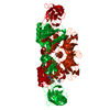
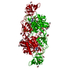
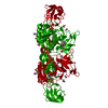
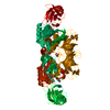
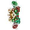
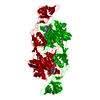
- PDB-6etf: The Structure of the Mo-insertase domain Cnx1E from Arabidopsis t... -
+ Open data

- Basic information
| Entry | Database: PDB / ID: 6etf | ||||||
|---|---|---|---|---|---|---|---|
| Title | The Structure of the Mo-insertase domain Cnx1E from Arabidopsis thaliana in complex with AMP and molybdate | ||||||
 Components Components | Molybdopterin biosynthesis protein CNX1 | ||||||
 Keywords Keywords | PLANT PROTEIN / Arabidopsis / Arabidopsis Proteins / Coenzymes / Metalloproteins / Catalytic Domain / Nucleotide Binding / Entropic Enzyme / Adenosine Monophosphate / molybdate / alternate binding / insertion mechanism | ||||||
| Function / homology |  Function and homology information Function and homology informationmolybdopterin adenylyltransferase activity / molybdopterin adenylyltransferase / nitrate reductase activity / molybdopterin molybdotransferase activity / molybdopterin molybdotransferase / auxin-activated signaling pathway / Mo-molybdopterin cofactor biosynthetic process / molybdenum ion binding / response to metal ion / ATP binding ...molybdopterin adenylyltransferase activity / molybdopterin adenylyltransferase / nitrate reductase activity / molybdopterin molybdotransferase activity / molybdopterin molybdotransferase / auxin-activated signaling pathway / Mo-molybdopterin cofactor biosynthetic process / molybdenum ion binding / response to metal ion / ATP binding / cytoplasm / cytosol Similarity search - Function | ||||||
| Biological species |  | ||||||
| Method |  X-RAY DIFFRACTION / SYNCHROTRON / MOLECULAR REPLACEMENT / Resolution: 1.781 Å X-RAY DIFFRACTION / SYNCHROTRON / MOLECULAR REPLACEMENT / Resolution: 1.781 Å | ||||||
 Authors Authors | Krausze, J. | ||||||
 Citation Citation | Journal: Biochem. J. / Year: 2018 Title: The functional principle of eukaryotic molybdenum insertases. Authors: Krausze, J. / Hercher, T.W. / Zwerschke, D. / Kirk, M.L. / Blankenfeldt, W. / Mendel, R.R. / Kruse, T. | ||||||
| History |
|
- Structure visualization
| Structure viewer | Molecule: MolmilJmol/JSmol |
|---|
- Downloads & links
-Download
| PDBx/mmCIF format | 6etf.cif.gz | 337.9 KB | Display | PDBx/mmCIF format |
|---|---|---|---|---|
| PDB format | pdb6etf.ent.gz | 275.4 KB | Display | PDB format |
| PDBx/mmJSON format | 6etf.json.gz | Tree view | PDBx/mmJSON format | |
| Others |  Other downloads Other downloads |
-Validation report
| Arichive directory | https://data.pdbj.org/pub/pdb/validation_reports/et/6etfftp://data.pdbj.org/pub/pdb/validation_reports/et/6etf | HTTPS FTP |
|---|
-Related structure data
| Related structure data |  6etdC  6ethC 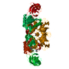 6gaxC  6gb0C 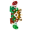 6gb4C  6gb9C  6gbcC 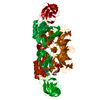 6gbfC  5g2rS S: Starting model for refinement C: citing same article ( |
|---|---|
| Similar structure data | |
| Other databases |
|


-Links
 PDBj
PDBj


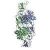
- Assembly
| Deposited unit | 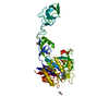
| ||||||||
|---|---|---|---|---|---|---|---|---|---|
| 1 | 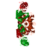
| ||||||||
| Unit cell |
| ||||||||
| Components on special symmetry positions |
|
-Components
-Protein , 1 types, 1 molecules A
| #1: Protein | Mass: 49763.094 Da / Num. of mol.: 1 Source method: isolated from a genetically manipulated source Source: (gene. exp.)  References: UniProt: Q39054, molybdopterin molybdotransferase, molybdopterin adenylyltransferase |
|---|
-Non-polymers , 5 types, 434 molecules 


| #2: Chemical | ChemComp-AMP /  Mass: 347.221 Da / Num. of mol.: 1 / Source method: obtained synthetically / Formula: C10H14N5O7P / Comment: AMP*YM Mass: 347.221 Da / Num. of mol.: 1 / Source method: obtained synthetically / Formula: C10H14N5O7P / Comment: AMP*YM | ||
|---|---|---|---|
| #3: Chemical | ChemComp-MG /  Mass: 24.305 Da / Num. of mol.: 1 / Source method: obtained synthetically / Formula: Mg Mass: 24.305 Da / Num. of mol.: 1 / Source method: obtained synthetically / Formula: Mg | ||
| #4: Chemical | ChemComp-MOO /  Mass: 159.938 Da / Num. of mol.: 1 / Source method: obtained synthetically / Formula: MoO4 / Feature type: SUBJECT OF INVESTIGATION Mass: 159.938 Da / Num. of mol.: 1 / Source method: obtained synthetically / Formula: MoO4 / Feature type: SUBJECT OF INVESTIGATION | ||
| #5: Chemical | ChemComp-EDO /  Mass: 62.068 Da / Num. of mol.: 4 / Source method: obtained synthetically / Formula: C2H6O2 Mass: 62.068 Da / Num. of mol.: 4 / Source method: obtained synthetically / Formula: C2H6O2#6: Water | ChemComp-HOH / | Mass: 18.015 Da / Num. of mol.: 427 / Source method: isolated from a natural source / Formula: H2O |
-Experimental details
-Experiment
| Experiment | Method: X-RAY DIFFRACTION / Number of used crystals: 1 |
|---|
- Sample preparation
| Crystal | Density Matthews: 2.72 Å3/Da / Density % sol: 54.82 % |
|---|---|
| Crystal grow | Temperature: 293 K / Method: vapor diffusion, sitting drop / pH: 7.5 Details: 20 % (v/v) 1,2-Ethanediol; 10 % (w/v) PEG 8000; 0.3 M MgCl2; 0.3 M CaCl2; 0.1 M Tris/BICINE, pH 7.4 |
-Data collection
| Diffraction | Mean temperature: 100 K |
|---|---|
| Diffraction source | Source: SYNCHROTRON / Site: BESSY  / Beamline: 14.1 / Wavelength: 1.77 Å / Beamline: 14.1 / Wavelength: 1.77 Å |
| Detector | Type: DECTRIS PILATUS3 6M / Detector: PIXEL / Date: Jun 3, 2017 / Details: mirrors |
| Radiation | Monochromator: SI111 double crystal with sagital bender / Protocol: SINGLE WAVELENGTH / Monochromatic (M) / Laue (L): M / Scattering type: x-ray |
| Radiation wavelength | Wavelength: 1.77 Å / Relative weight: 1 |
| Reflection | Resolution: 1.781→45.072 Å / Num. obs: 34559 / % possible obs: 65.61 % / Observed criterion σ(I): -3 / Redundancy: 15.38 % / Biso Wilson estimate: 36.87 Å2 / CC1/2: 0.9997 / Rmerge(I) obs: 0.054 / Rpim(I) all: 0.014 / Rrim(I) all: 0.056 / Net I/σ(I): 26.558 |
| Reflection shell | Resolution: 1.781→1.963 Å / Redundancy: 9.72 % / Rmerge(I) obs: 1.339 / Mean I/σ(I) obs: 1.527 / Num. unique obs: 1728 / CC1/2: 0.719 / Rpim(I) all: 0.443 / Rrim(I) all: 1.414 / % possible all: 13.18 |
- Processing
| Software |
| ||||||||||||||||||||||||||||||||||||||||||||||||||||||||||||||||||||||||||||||||||||||||||||||||||||||||||||||||||
|---|---|---|---|---|---|---|---|---|---|---|---|---|---|---|---|---|---|---|---|---|---|---|---|---|---|---|---|---|---|---|---|---|---|---|---|---|---|---|---|---|---|---|---|---|---|---|---|---|---|---|---|---|---|---|---|---|---|---|---|---|---|---|---|---|---|---|---|---|---|---|---|---|---|---|---|---|---|---|---|---|---|---|---|---|---|---|---|---|---|---|---|---|---|---|---|---|---|---|---|---|---|---|---|---|---|---|---|---|---|---|---|---|---|---|---|
| Refinement | Method to determine structure: MOLECULAR REPLACEMENT Starting model: 5G2R Resolution: 1.781→33.14 Å / Cor.coef. Fo:Fc: 0.956 / Cor.coef. Fo:Fc free: 0.943 / SU R Cruickshank DPI: 0.258 / Cross valid method: THROUGHOUT / σ(F): 0 / SU R Blow DPI: 0.166 / SU Rfree Blow DPI: 0.15 / SU Rfree Cruickshank DPI: 0.144
| ||||||||||||||||||||||||||||||||||||||||||||||||||||||||||||||||||||||||||||||||||||||||||||||||||||||||||||||||||
| Displacement parameters | Biso mean: 38.46 Å2
| ||||||||||||||||||||||||||||||||||||||||||||||||||||||||||||||||||||||||||||||||||||||||||||||||||||||||||||||||||
| Refine analyze | Luzzati coordinate error obs: 0.24 Å | ||||||||||||||||||||||||||||||||||||||||||||||||||||||||||||||||||||||||||||||||||||||||||||||||||||||||||||||||||
| Refinement step | Cycle: 1 / Resolution: 1.781→33.14 Å
| ||||||||||||||||||||||||||||||||||||||||||||||||||||||||||||||||||||||||||||||||||||||||||||||||||||||||||||||||||
| Refine LS restraints |
| ||||||||||||||||||||||||||||||||||||||||||||||||||||||||||||||||||||||||||||||||||||||||||||||||||||||||||||||||||
| LS refinement shell | Resolution: 1.78→1.84 Å / Total num. of bins used: 17
| ||||||||||||||||||||||||||||||||||||||||||||||||||||||||||||||||||||||||||||||||||||||||||||||||||||||||||||||||||
| Refinement TLS params. | Method: refined / Refine-ID: X-RAY DIFFRACTION
| ||||||||||||||||||||||||||||||||||||||||||||||||||||||||||||||||||||||||||||||||||||||||||||||||||||||||||||||||||
| Refinement TLS group |
|
