 Movie
Movie Controller

[English] 日本語
 Yorodumi
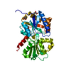
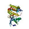
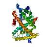
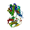
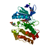
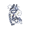
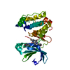
- PDB-6esv: Structure of the phosphate-bound form of AioX from Rhizobium sp. ... -
+ Open data

- Basic information
| Entry | Database: PDB / ID: 6esv | |||||||||
|---|---|---|---|---|---|---|---|---|---|---|
| Title | Structure of the phosphate-bound form of AioX from Rhizobium sp. str. NT-26 | |||||||||
 Components Components | Putative periplasmic phosphite-binding-like protein (Pbl) PtxB-like protein designated AioX | |||||||||
 Keywords Keywords | SIGNALING PROTEIN / Periplasmic-binding protein / Arsenic / arsenite-binding / Rhizobium NT-26 | |||||||||
| Function / homology | ABC transporter, phosphonate, periplasmic substrate-binding protein / PHOSPHATE ION / Putative periplasmic phosphite-binding-like protein (Pbl) PtxB-like protein designated AioX Function and homology information Function and homology information | |||||||||
| Biological species |  Rhizobium sp. NT-26 (bacteria) Rhizobium sp. NT-26 (bacteria) | |||||||||
| Method |  X-RAY DIFFRACTION / SYNCHROTRON / SAD / Resolution: 1.78 Å X-RAY DIFFRACTION / SYNCHROTRON / SAD / Resolution: 1.78 Å | |||||||||
 Authors Authors | Djordjevic, S. / Badilla, C. / Cole, A. / Santini, J. | |||||||||
| Funding support |  Chile, Chile,  United Kingdom, 2items United Kingdom, 2items
| |||||||||
 Citation Citation | Journal: Sci Rep / Year: 2018 Title: A new family of periplasmic-binding proteins that sense arsenic oxyanions. Authors: Badilla, C. / Osborne, T.H. / Cole, A. / Watson, C. / Djordjevic, S. / Santini, J.M. | |||||||||
| History |
|
- Structure visualization
| Structure viewer | Molecule: MolmilJmol/JSmol |
|---|
- Downloads & links
-Download
| PDBx/mmCIF format | 6esv.cif.gz | 70.2 KB | Display | PDBx/mmCIF format |
|---|---|---|---|---|
| PDB format | pdb6esv.ent.gz | 50.2 KB | Display | PDB format |
| PDBx/mmJSON format | 6esv.json.gz | Tree view | PDBx/mmJSON format | |
| Others |  Other downloads Other downloads |
-Validation report
| Arichive directory | https://data.pdbj.org/pub/pdb/validation_reports/es/6esvftp://data.pdbj.org/pub/pdb/validation_reports/es/6esv | HTTPS FTP |
|---|
-Related structure data


-Links
 PDBj
PDBj- Assembly
| Deposited unit | 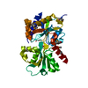
| ||||||||
|---|---|---|---|---|---|---|---|---|---|
| 1 |
| ||||||||
| Unit cell |
|
-Components
| #1: Protein | Mass: 31426.639 Da / Num. of mol.: 1 Source method: isolated from a genetically manipulated source Details: This is a Se-Met labeled protein which is why MET residues are replaced with MSE. Two residues, K230 and R173 (or K250 and R193 as numbered in the PDB file to reflect the protein numbering ...Details: This is a Se-Met labeled protein which is why MET residues are replaced with MSE. Two residues, K230 and R173 (or K250 and R193 as numbered in the PDB file to reflect the protein numbering after the transport signal peptide was removed) were modeled as Alanines due to the disorder for the side chain. Similarly, residues 37 and 38 (57 and 58) were not modeled due to disorder. Source: (gene. exp.) Rhizobium sp. NT-26 (bacteria) / Gene: aioX, NT26_p10026 / Production host: |
|---|---|
| #2: Chemical | ChemComp-PO4 /   Mass: 94.971 Da / Num. of mol.: 1 / Source method: obtained synthetically / Formula: PO4 Mass: 94.971 Da / Num. of mol.: 1 / Source method: obtained synthetically / Formula: PO4 |
| #3: Water | ChemComp-HOH /  Mass: 18.015 Da / Num. of mol.: 127 / Source method: isolated from a natural source / Formula: H2O Mass: 18.015 Da / Num. of mol.: 127 / Source method: isolated from a natural source / Formula: H2O |
| Has protein modification | Y |
-Experimental details
-Experiment
| Experiment | Method: X-RAY DIFFRACTION / Number of used crystals: 1 |
|---|
- Sample preparation
| Crystal | Density Matthews: 2.99 Å3/Da / Density % sol: 58.85 % |
|---|---|
| Crystal grow | Temperature: 289.15 K / Method: vapor diffusion, hanging drop / pH: 7.2 / Details: 0.49 M NaH2PO4 and 0.91 M K2HPO4 |
-Data collection
| Diffraction | Mean temperature: 100 K |
|---|---|
| Diffraction source | Source: SYNCHROTRON / Site: Diamond / Beamline: I04 / Wavelength: 0.978 Å |
| Detector | Type: DECTRIS PILATUS 6M-F / Detector: PIXEL / Date: Apr 13, 2015 |
| Radiation | Protocol: SINGLE WAVELENGTH / Monochromatic (M) / Laue (L): M / Scattering type: x-ray |
| Radiation wavelength | Wavelength: 0.978 Å / Relative weight: 1 |
| Reflection | Resolution: 1.78→88.07 Å / Num. obs: 36718 / % possible obs: 99.8 % / Redundancy: 16.4 % / Net I/σ(I): 19.3 |
- Processing
| Software |
| ||||||||||||||||||||||||||||||||||||||||||||||||||||||||||||||||||||||||||||||||||||||||||||||||||||||||||||||||||||||||||||||||||||||||||||||||||||||||||||||||||||||||||||||||||||||
|---|---|---|---|---|---|---|---|---|---|---|---|---|---|---|---|---|---|---|---|---|---|---|---|---|---|---|---|---|---|---|---|---|---|---|---|---|---|---|---|---|---|---|---|---|---|---|---|---|---|---|---|---|---|---|---|---|---|---|---|---|---|---|---|---|---|---|---|---|---|---|---|---|---|---|---|---|---|---|---|---|---|---|---|---|---|---|---|---|---|---|---|---|---|---|---|---|---|---|---|---|---|---|---|---|---|---|---|---|---|---|---|---|---|---|---|---|---|---|---|---|---|---|---|---|---|---|---|---|---|---|---|---|---|---|---|---|---|---|---|---|---|---|---|---|---|---|---|---|---|---|---|---|---|---|---|---|---|---|---|---|---|---|---|---|---|---|---|---|---|---|---|---|---|---|---|---|---|---|---|---|---|---|---|
| Refinement | Method to determine structure: SAD / Resolution: 1.78→88.07 Å / Cor.coef. Fo:Fc: 0.959 / Cor.coef. Fo:Fc free: 0.947 / SU B: 2.67 / SU ML: 0.08 / Cross valid method: THROUGHOUT / ESU R: 0.104 / ESU R Free: 0.103 / Details: HYDROGENS HAVE BEEN ADDED IN THE RIDING POSITIONS
| ||||||||||||||||||||||||||||||||||||||||||||||||||||||||||||||||||||||||||||||||||||||||||||||||||||||||||||||||||||||||||||||||||||||||||||||||||||||||||||||||||||||||||||||||||||||
| Solvent computation | Ion probe radii: 0.8 Å / Shrinkage radii: 0.8 Å / VDW probe radii: 1.2 Å | ||||||||||||||||||||||||||||||||||||||||||||||||||||||||||||||||||||||||||||||||||||||||||||||||||||||||||||||||||||||||||||||||||||||||||||||||||||||||||||||||||||||||||||||||||||||
| Displacement parameters | Biso mean: 31.742 Å2
| ||||||||||||||||||||||||||||||||||||||||||||||||||||||||||||||||||||||||||||||||||||||||||||||||||||||||||||||||||||||||||||||||||||||||||||||||||||||||||||||||||||||||||||||||||||||
| Refinement step | Cycle: 1 / Resolution: 1.78→88.07 Å
| ||||||||||||||||||||||||||||||||||||||||||||||||||||||||||||||||||||||||||||||||||||||||||||||||||||||||||||||||||||||||||||||||||||||||||||||||||||||||||||||||||||||||||||||||||||||
| Refine LS restraints |
|