 Movie
Movie Controller


+ Open data

- Basic information
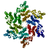
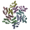
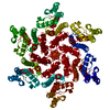
| Entry | Database: PDB / ID: 6erm | ||||||
|---|---|---|---|---|---|---|---|
| Title | HIV Hexamer with ligand | ||||||
 Components Components | Gag polyprotein | ||||||
 Keywords Keywords | VIRAL PROTEIN / HIV | ||||||
| Function / homology |  Function and homology information Function and homology informationviral nucleocapsid / host cell cytoplasm / viral translational frameshifting / host cell nucleus / virion membrane / structural molecule activity / RNA binding / zinc ion binding / ATP binding Similarity search - Function | ||||||
| Biological species |   Human immunodeficiency virus 1 Human immunodeficiency virus 1 | ||||||
| Method |  X-RAY DIFFRACTION / SYNCHROTRON / MOLECULAR REPLACEMENT / Resolution: 2 Å X-RAY DIFFRACTION / SYNCHROTRON / MOLECULAR REPLACEMENT / Resolution: 2 Å | ||||||
 Authors Authors | James, L.C. | ||||||
 Citation Citation | Journal: To Be Published Title: HIV hexamer Authors: James, L.C. | ||||||
| History |
|
- Structure visualization
| Structure viewer | Molecule: MolmilJmol/JSmol |
|---|
- Downloads & links
-Download
| PDBx/mmCIF format | 6erm.cif.gz | 60.8 KB | Display | PDBx/mmCIF format |
|---|---|---|---|---|
| PDB format | pdb6erm.ent.gz | 42.3 KB | Display | PDB format |
| PDBx/mmJSON format | 6erm.json.gz | Tree view | PDBx/mmJSON format | |
| Others |  Other downloads Other downloads |
-Validation report
| Summary document | 6erm_validation.pdf.gz | 676 KB | Display | wwPDB validaton report |
|---|---|---|---|---|
| Full document | 6erm_full_validation.pdf.gz | 678.2 KB | Display | |
| Data in XML | 6erm_validation.xml.gz | 14.5 KB | Display | |
| Data in CIF | 6erm_validation.cif.gz | 20.4 KB | Display | |
| Arichive directory | https://data.pdbj.org/pub/pdb/validation_reports/er/6ermftp://data.pdbj.org/pub/pdb/validation_reports/er/6erm | HTTPS FTP |
-Related structure data
| Related structure data | 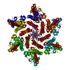 6ernC  6r6qC  6r8cC  5hgmS S: Starting model for refinement C: citing same article ( |
|---|---|
| Similar structure data |


-Links
 PDBj
PDBj
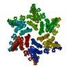
- Assembly
| Deposited unit | 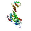
| ||||||||||||||||||
|---|---|---|---|---|---|---|---|---|---|---|---|---|---|---|---|---|---|---|---|
| 1 | x 6
| ||||||||||||||||||
| Unit cell |
| ||||||||||||||||||
| Components on special symmetry positions |
|
-Components
| #1: Protein | Mass: 24504.178 Da / Num. of mol.: 1 Source method: isolated from a genetically manipulated source Source: (gene. exp.) Human immunodeficiency virus 1 / Gene: gag / Production host:  |
|---|---|
| #2: Chemical | ChemComp-AZT /   Mass: 507.181 Da / Num. of mol.: 1 / Source method: obtained synthetically / Formula: C10H16N5O13P3 Mass: 507.181 Da / Num. of mol.: 1 / Source method: obtained synthetically / Formula: C10H16N5O13P3 |
| #3: Water | ChemComp-HOH /  Mass: 18.015 Da / Num. of mol.: 210 / Source method: isolated from a natural source / Formula: H2O Mass: 18.015 Da / Num. of mol.: 210 / Source method: isolated from a natural source / Formula: H2O |
| Has protein modification | Y |
-Experimental details
-Experiment
| Experiment | Method: X-RAY DIFFRACTION / Number of used crystals: 1 |
|---|
- Sample preparation
| Crystal | Density Matthews: 2.98 Å3/Da / Density % sol: 58.76 % |
|---|---|
| Crystal grow | Temperature: 277 K / Method: batch mode / Details: PEG4K |
-Data collection
| Diffraction | Mean temperature: 100 K |
|---|---|
| Diffraction source | Source: SYNCHROTRON / Site: Diamond  / Beamline: I02 / Wavelength: 1 Å / Beamline: I02 / Wavelength: 1 Å |
| Detector | Type: OXFORD RUBY CCD / Detector: CCD / Date: Jan 3, 2017 |
| Radiation | Protocol: SINGLE WAVELENGTH / Monochromatic (M) / Laue (L): M / Scattering type: x-ray |
| Radiation wavelength | Wavelength: 1 Å / Relative weight: 1 |
| Reflection | Resolution: 2→78 Å / Num. obs: 18217 / % possible obs: 100 % / Redundancy: 9.3 % / Net I/σ(I): 10.8 |
| Reflection shell | Resolution: 2→2.03 Å |
- Processing
| Software |
| ||||||||||||||||||||||||||||||||||||||||||||||||||||||||||||||||||||||||||||||||||||||||||||||||||||||||||||||||||||||||||||||||||||||||||||||||||||||||||||||||||||||||||||||||||||||
|---|---|---|---|---|---|---|---|---|---|---|---|---|---|---|---|---|---|---|---|---|---|---|---|---|---|---|---|---|---|---|---|---|---|---|---|---|---|---|---|---|---|---|---|---|---|---|---|---|---|---|---|---|---|---|---|---|---|---|---|---|---|---|---|---|---|---|---|---|---|---|---|---|---|---|---|---|---|---|---|---|---|---|---|---|---|---|---|---|---|---|---|---|---|---|---|---|---|---|---|---|---|---|---|---|---|---|---|---|---|---|---|---|---|---|---|---|---|---|---|---|---|---|---|---|---|---|---|---|---|---|---|---|---|---|---|---|---|---|---|---|---|---|---|---|---|---|---|---|---|---|---|---|---|---|---|---|---|---|---|---|---|---|---|---|---|---|---|---|---|---|---|---|---|---|---|---|---|---|---|---|---|---|---|
| Refinement | Method to determine structure: MOLECULAR REPLACEMENT Starting model: 5HGM Resolution: 2→78 Å / Cor.coef. Fo:Fc: 0.941 / Cor.coef. Fo:Fc free: 0.906 / SU B: 3.466 / SU ML: 0.099 / Cross valid method: THROUGHOUT / ESU R: 0.155 / ESU R Free: 0.154 / Details: HYDROGENS HAVE BEEN ADDED IN THE RIDING POSITIONS
| ||||||||||||||||||||||||||||||||||||||||||||||||||||||||||||||||||||||||||||||||||||||||||||||||||||||||||||||||||||||||||||||||||||||||||||||||||||||||||||||||||||||||||||||||||||||
| Solvent computation | Ion probe radii: 0.8 Å / Shrinkage radii: 0.8 Å / VDW probe radii: 1.2 Å | ||||||||||||||||||||||||||||||||||||||||||||||||||||||||||||||||||||||||||||||||||||||||||||||||||||||||||||||||||||||||||||||||||||||||||||||||||||||||||||||||||||||||||||||||||||||
| Displacement parameters | Biso mean: 29.017 Å2
| ||||||||||||||||||||||||||||||||||||||||||||||||||||||||||||||||||||||||||||||||||||||||||||||||||||||||||||||||||||||||||||||||||||||||||||||||||||||||||||||||||||||||||||||||||||||
| Refinement step | Cycle: 1 / Resolution: 2→78 Å
| ||||||||||||||||||||||||||||||||||||||||||||||||||||||||||||||||||||||||||||||||||||||||||||||||||||||||||||||||||||||||||||||||||||||||||||||||||||||||||||||||||||||||||||||||||||||
| Refine LS restraints |
|