 Movie
Movie Controller

[English] 日本語
 Yorodumi
- PDB-6en3: Crystal structure of full length EndoS from Streptococcus pyogene... -
+ Open data

- Basic information
| Entry | Database: PDB / ID: 6en3 | |||||||||
|---|---|---|---|---|---|---|---|---|---|---|
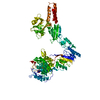
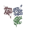
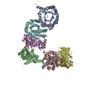
| Title | Crystal structure of full length EndoS from Streptococcus pyogenes in complex with G2 oligosaccharide. | |||||||||
 Components Components | Endo-beta-N-acetylglucosaminidase F2,Multifunctional-autoprocessing repeats-in-toxin | |||||||||
 Keywords Keywords | HYDROLASE / endoglycosidase/immunoglobulin/carbohydrate/endo-beta-N-acetylglucos aminidase | |||||||||
| Function / homology |  Function and homology information Function and homology informationCoA-dependent peptidyl-lysine N6-palmitoyltransferase activity / Ligases; Forming carbon-nitrogen bonds; Acid-amino-acid ligases (peptide synthases) / host cell cytosol / ligase activity / hydrolase activity, hydrolyzing O-glycosyl compounds / cysteine-type peptidase activity / Transferases; Acyltransferases; Transferring groups other than aminoacyl groups / actin filament organization / toxin activity / carbohydrate metabolic process ...CoA-dependent peptidyl-lysine N6-palmitoyltransferase activity / Ligases; Forming carbon-nitrogen bonds; Acid-amino-acid ligases (peptide synthases) / host cell cytosol / ligase activity / hydrolase activity, hydrolyzing O-glycosyl compounds / cysteine-type peptidase activity / Transferases; Acyltransferases; Transferring groups other than aminoacyl groups / actin filament organization / toxin activity / carbohydrate metabolic process / Hydrolases; Acting on peptide bonds (peptidases); Cysteine endopeptidases / lipid binding / host cell plasma membrane / proteolysis / extracellular region / ATP binding / metal ion binding Similarity search - Function | |||||||||
| Biological species |  Streptococcus pyogenes (bacteria) Streptococcus pyogenes (bacteria) Vibrio cholerae (bacteria) Vibrio cholerae (bacteria) | |||||||||
| Method |  X-RAY DIFFRACTION / SYNCHROTRON / MOLECULAR REPLACEMENT / Resolution: 2.903 Å X-RAY DIFFRACTION / SYNCHROTRON / MOLECULAR REPLACEMENT / Resolution: 2.903 Å | |||||||||
 Authors Authors | Trastoy, B. / Klontz, E.H. / Orwenyo, J. / Marina, A. / Wang, L.X. / Sundberg, E.J. / Guerin, M.E. | |||||||||
 Citation Citation | Journal: Nat Commun / Year: 2018 Title: Structural basis for the recognition of complex-type N-glycans by Endoglycosidase S. Authors: Trastoy, B. / Klontz, E. / Orwenyo, J. / Marina, A. / Wang, L.X. / Sundberg, E.J. / Guerin, M.E. | |||||||||
| History |
|
- Structure visualization
| Structure viewer | Molecule: MolmilJmol/JSmol |
|---|
- Downloads & links
-Download
| PDBx/mmCIF format | 6en3.cif.gz | 392.9 KB | Display | PDBx/mmCIF format |
|---|---|---|---|---|
| PDB format | pdb6en3.ent.gz | 316.4 KB | Display | PDB format |
| PDBx/mmJSON format | 6en3.json.gz | Tree view | PDBx/mmJSON format | |
| Others |  Other downloads Other downloads |
-Validation report
| Arichive directory | https://data.pdbj.org/pub/pdb/validation_reports/en/6en3ftp://data.pdbj.org/pub/pdb/validation_reports/en/6en3 | HTTPS FTP |
|---|
-Related structure data
| Related structure data | 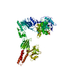 4nuzS S: Starting model for refinement |
|---|---|
| Similar structure data |


-Links
 PDBj
PDBj


- Assembly
| Deposited unit | 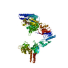
| ||||||||
|---|---|---|---|---|---|---|---|---|---|
| 1 |
| ||||||||
| Unit cell |
|
-Components
| #1: Protein | Mass: 133360.219 Da / Num. of mol.: 1 / Mutation: D233A, L235E Source method: isolated from a genetically manipulated source Source: (gene. exp.) Streptococcus pyogenes (bacteria), (gene. exp.) Vibrio cholerae (bacteria)Gene: endoS, M1GAS476_1618, rtxA, rtx, VC_1451 / Production host: References: UniProt: J7M8R4, UniProt: Q9KS12, Hydrolases; Acting on peptide bonds (peptidases); Cysteine endopeptidases, Ligases; Forming carbon-nitrogen bonds; Acid-amino-acid ligases (peptide synthases) |
|---|---|
| #2: Polysaccharide | beta-D-galactopyranose-(1-4)-2-acetamido-2-deoxy-beta-D-glucopyranose-(1-2)-alpha-D-mannopyranose- ...beta-D-galactopyranose-(1-4)-2-acetamido-2-deoxy-beta-D-glucopyranose-(1-2)-alpha-D-mannopyranose-(1-3)-[beta-D-galactopyranose-(1-4)-2-acetamido-2-deoxy-beta-D-glucopyranose-(1-2)-alpha-D-mannopyranose-(1-6)]beta-D-mannopyranose-(1-4)-2-acetamido-2-deoxy-beta-D-glucopyranose Source method: isolated from a genetically manipulated source |
| #3: Chemical | ChemComp-CA /   Mass: 40.078 Da / Num. of mol.: 1 / Source method: obtained synthetically / Formula: Ca Mass: 40.078 Da / Num. of mol.: 1 / Source method: obtained synthetically / Formula: Ca |
| #4: Chemical | ChemComp-NI /   Mass: 58.693 Da / Num. of mol.: 1 Mass: 58.693 Da / Num. of mol.: 1Source method: isolated from a genetically manipulated source Formula: Ni |
| #5: Water | ChemComp-HOH /  Mass: 18.015 Da / Num. of mol.: 7 / Source method: isolated from a natural source / Formula: H2O Mass: 18.015 Da / Num. of mol.: 7 / Source method: isolated from a natural source / Formula: H2O |
-Experimental details
-Experiment
| Experiment | Method: X-RAY DIFFRACTION / Number of used crystals: 1 |
|---|
- Sample preparation
| Crystal | Density Matthews: 2.56 Å3/Da / Density % sol: 52.04 % |
|---|---|
| Crystal grow | Temperature: 298 K / Method: vapor diffusion, sitting drop / pH: 7.5 Details: 100 mM Sodium HEPES/MOPS pH 7.5, 100 mM aminoacids (L-Na-Glutamate, alanine (racemic), glycine, lysine HCL (racemic), serine (racemic)), 20% (w/v) PEG 500 MME and 10% (w/v) PEG 20,000 |
-Data collection
| Diffraction | Mean temperature: 100 K |
|---|---|
| Diffraction source | Source: SYNCHROTRON / Site: SOLEIL  / Beamline: PROXIMA 2 / Wavelength: 0.980105 Å / Beamline: PROXIMA 2 / Wavelength: 0.980105 Å |
| Detector | Type: DECTRIS EIGER X 9M / Detector: PIXEL / Date: Dec 2, 2016 |
| Radiation | Protocol: SINGLE WAVELENGTH / Monochromatic (M) / Laue (L): M / Scattering type: x-ray |
| Radiation wavelength | Wavelength: 0.980105 Å / Relative weight: 1 |
| Reflection | Resolution: 2.9→45.04 Å / Num. obs: 118762 / % possible obs: 98.8 % / Redundancy: 4 % / CC1/2: 0.999 / Rmerge(I) obs: 0.096 / Net I/σ(I): 10.6 |
| Reflection shell | Resolution: 2.9→3.007 Å / Redundancy: 4.1 % / Rmerge(I) obs: 0.781 / CC1/2: 0.852 / % possible all: 100 |
- Processing
| Software |
| ||||||||||||||||||||||||||||||||||||||||||||||||||||||||||||||||||||||||||||||||||||||||||||||||||||||||||||||||||||||||||||||||||||||||||||||||||||||||||
|---|---|---|---|---|---|---|---|---|---|---|---|---|---|---|---|---|---|---|---|---|---|---|---|---|---|---|---|---|---|---|---|---|---|---|---|---|---|---|---|---|---|---|---|---|---|---|---|---|---|---|---|---|---|---|---|---|---|---|---|---|---|---|---|---|---|---|---|---|---|---|---|---|---|---|---|---|---|---|---|---|---|---|---|---|---|---|---|---|---|---|---|---|---|---|---|---|---|---|---|---|---|---|---|---|---|---|---|---|---|---|---|---|---|---|---|---|---|---|---|---|---|---|---|---|---|---|---|---|---|---|---|---|---|---|---|---|---|---|---|---|---|---|---|---|---|---|---|---|---|---|---|---|---|---|---|
| Refinement | Method to determine structure: MOLECULAR REPLACEMENT Starting model: 4NUZ Resolution: 2.903→45.04 Å / SU ML: 0.44 / Cross valid method: FREE R-VALUE / σ(F): 0.63 / Phase error: 26.24 / Stereochemistry target values: ML
| ||||||||||||||||||||||||||||||||||||||||||||||||||||||||||||||||||||||||||||||||||||||||||||||||||||||||||||||||||||||||||||||||||||||||||||||||||||||||||
| Solvent computation | Shrinkage radii: 0.9 Å / VDW probe radii: 1.11 Å / Solvent model: FLAT BULK SOLVENT MODEL | ||||||||||||||||||||||||||||||||||||||||||||||||||||||||||||||||||||||||||||||||||||||||||||||||||||||||||||||||||||||||||||||||||||||||||||||||||||||||||
| Refinement step | Cycle: LAST / Resolution: 2.903→45.04 Å
| ||||||||||||||||||||||||||||||||||||||||||||||||||||||||||||||||||||||||||||||||||||||||||||||||||||||||||||||||||||||||||||||||||||||||||||||||||||||||||
| Refine LS restraints |
| ||||||||||||||||||||||||||||||||||||||||||||||||||||||||||||||||||||||||||||||||||||||||||||||||||||||||||||||||||||||||||||||||||||||||||||||||||||||||||
| LS refinement shell |
| ||||||||||||||||||||||||||||||||||||||||||||||||||||||||||||||||||||||||||||||||||||||||||||||||||||||||||||||||||||||||||||||||||||||||||||||||||||||||||
| Refinement TLS params. | Method: refined / Refine-ID: X-RAY DIFFRACTION
| ||||||||||||||||||||||||||||||||||||||||||||||||||||||||||||||||||||||||||||||||||||||||||||||||||||||||||||||||||||||||||||||||||||||||||||||||||||||||||
| Refinement TLS group |
|
