 Movie
Movie Controller

[English] 日本語
 Yorodumi
- PDB-6ejq: Crystal structure of a C-terminally truncated small terminase pro... -
+ Open data

- Basic information
| Entry | Database: PDB / ID: 6ejq | |||||||||
|---|---|---|---|---|---|---|---|---|---|---|
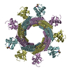
| Title | Crystal structure of a C-terminally truncated small terminase protein from the thermophilic bacteriophage G20c | |||||||||
 Components Components | Terminase small subunit | |||||||||
 Keywords Keywords | VIRAL PROTEIN / Small terminase | |||||||||
| Function / homology | : / : / Phage G20C small terminase, N-terminal domain / Phage G20C small terminase C-terminal domain / DNA binding / Terminase, small subunit Function and homology information Function and homology information | |||||||||
| Biological species |   Thermus phage G20c (virus) Thermus phage G20c (virus) | |||||||||
| Method |  X-RAY DIFFRACTION / SYNCHROTRON / MOLECULAR REPLACEMENT / Resolution: 2.3 Å X-RAY DIFFRACTION / SYNCHROTRON / MOLECULAR REPLACEMENT / Resolution: 2.3 Å | |||||||||
 Authors Authors | Xu, R.G. / Loredo-Varela, J. / Jenkins, H.T. / Antson, A.A. | |||||||||
| Funding support |  United Kingdom, 2items United Kingdom, 2items
| |||||||||
 Citation Citation | Journal: To Be Published Title: Crystal structure of a C-terminally truncated small terminase protein from the thermophilic bacteriophage G20c Authors: Xu, R.G. / Loredo-Varela, J. / Jenkins, H.T. / Antson, A.A. | |||||||||
| History |
|
- Structure visualization
| Structure viewer | Molecule: MolmilJmol/JSmol |
|---|
- Downloads & links
-Download
| PDBx/mmCIF format | 6ejq.cif.gz | 241.1 KB | Display | PDBx/mmCIF format |
|---|---|---|---|---|
| PDB format | pdb6ejq.ent.gz | 196.8 KB | Display | PDB format |
| PDBx/mmJSON format | 6ejq.json.gz | Tree view | PDBx/mmJSON format | |
| Others |  Other downloads Other downloads |
-Validation report
| Arichive directory | https://data.pdbj.org/pub/pdb/validation_reports/ej/6ejqftp://data.pdbj.org/pub/pdb/validation_reports/ej/6ejq | HTTPS FTP |
|---|
-Related structure data
| Related structure data | 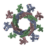 4xvnS S: Starting model for refinement |
|---|---|
| Similar structure data |


-Links
 PDBj
PDBj- Assembly
| Deposited unit | 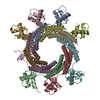
| ||||||||
|---|---|---|---|---|---|---|---|---|---|
| 1 |
| ||||||||
| Unit cell |
|
-Components
| #1: Protein | Mass: 15805.306 Da / Num. of mol.: 9 Source method: isolated from a genetically manipulated source Details: Two helices of chain E haven't been model due to poor density. Source: (gene. exp.) Thermus phage G20c (virus) / Gene: G20c_79 / Production host:  #2: Water | ChemComp-HOH / |  Mass: 18.015 Da / Num. of mol.: 653 / Source method: isolated from a natural source / Formula: H2O Mass: 18.015 Da / Num. of mol.: 653 / Source method: isolated from a natural source / Formula: H2O |
|---|
-Experimental details
-Experiment
| Experiment | Method: X-RAY DIFFRACTION / Number of used crystals: 1 |
|---|
- Sample preparation
| Crystal | Density Matthews: 3.6 Å3/Da / Density % sol: 65.79 % |
|---|---|
| Crystal grow | Temperature: 293 K / Method: vapor diffusion, sitting drop / pH: 7 / Details: 35% v/v Tacsimate pH 7.0 |
-Data collection
| Diffraction | Mean temperature: 273 K | ||||||||||||||||||||||||||||||
|---|---|---|---|---|---|---|---|---|---|---|---|---|---|---|---|---|---|---|---|---|---|---|---|---|---|---|---|---|---|---|---|
| Diffraction source | Source: SYNCHROTRON / Site: Diamond / Beamline: I04 / Wavelength: 0.9795 Å | ||||||||||||||||||||||||||||||
| Detector | Type: DECTRIS PILATUS 6M / Detector: PIXEL / Date: Jan 30, 2015 | ||||||||||||||||||||||||||||||
| Radiation | Protocol: SINGLE WAVELENGTH / Monochromatic (M) / Laue (L): M / Scattering type: x-ray | ||||||||||||||||||||||||||||||
| Radiation wavelength | Wavelength: 0.9795 Å / Relative weight: 1 | ||||||||||||||||||||||||||||||
| Reflection | Resolution: 2.3→49.47 Å / Num. obs: 88169 / % possible obs: 100 % / Redundancy: 13.5 % / CC1/2: 0.999 / Rmerge(I) obs: 0.126 / Rpim(I) all: 0.035 / Rrim(I) all: 0.131 / Net I/σ(I): 16.1 / Num. measured all: 1187820 / Scaling rejects: 0 | ||||||||||||||||||||||||||||||
| Reflection shell | Diffraction-ID: 1
|
- Processing
| Software |
| ||||||||||||||||||||||||||||||||||||||||||||||||||||||||||||
|---|---|---|---|---|---|---|---|---|---|---|---|---|---|---|---|---|---|---|---|---|---|---|---|---|---|---|---|---|---|---|---|---|---|---|---|---|---|---|---|---|---|---|---|---|---|---|---|---|---|---|---|---|---|---|---|---|---|---|---|---|---|
| Refinement | Method to determine structure: MOLECULAR REPLACEMENT Starting model: 4XVN Resolution: 2.3→49.47 Å / Cor.coef. Fo:Fc: 0.95 / Cor.coef. Fo:Fc free: 0.939 / SU B: 6.596 / SU ML: 0.153 / Cross valid method: THROUGHOUT / σ(F): 0 / ESU R: 0.226 / ESU R Free: 0.189 / Stereochemistry target values: MAXIMUM LIKELIHOOD Details: HYDROGENS HAVE BEEN ADDED IN THE RIDING POSITIONS U VALUES : REFINED INDIVIDUALLY
| ||||||||||||||||||||||||||||||||||||||||||||||||||||||||||||
| Solvent computation | Ion probe radii: 0.8 Å / Shrinkage radii: 0.8 Å / VDW probe radii: 1.2 Å / Solvent model: MASK | ||||||||||||||||||||||||||||||||||||||||||||||||||||||||||||
| Displacement parameters | Biso max: 142.1 Å2 / Biso mean: 56.049 Å2 / Biso min: 23.52 Å2
| ||||||||||||||||||||||||||||||||||||||||||||||||||||||||||||
| Refinement step | Cycle: final / Resolution: 2.3→49.47 Å
| ||||||||||||||||||||||||||||||||||||||||||||||||||||||||||||
| Refine LS restraints |
| ||||||||||||||||||||||||||||||||||||||||||||||||||||||||||||
| LS refinement shell | Resolution: 2.3→2.36 Å / Rfactor Rfree error: 0 / Total num. of bins used: 20
|
