 Movie
Movie Controller

[English] 日本語
 Yorodumi
- PDB-6ej7: Human Xylosyltransferase 1 in complex with UDP-xylose and peptide... -
+ Open data

- Basic information
| Entry | Database: PDB / ID: 6ej7 | ||||||
|---|---|---|---|---|---|---|---|
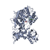
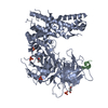
| Title | Human Xylosyltransferase 1 in complex with UDP-xylose and peptide QEEEGAGGGQGG | ||||||
 Components Components |
| ||||||
 Keywords Keywords | TRANSFERASE / Proteoglycan / Glycosyltransferase / Golgi / Xylosyltransferase | ||||||
| Function / homology |  Function and homology information Function and homology informationprotein xylosyltransferase / protein xylosyltransferase activity / Oxidoreductases; Acting on NADH or NADPH; With a heme protein as acceptor / calcium oxalate binding / glycosaminoglycan-protein linkage region biosynthetic process / Golgi cis cisterna / Glycosaminoglycan-protein linkage region biosynthesis / glycosaminoglycan biosynthetic process / proteoglycan biosynthetic process / chondroitin sulfate proteoglycan biosynthetic process ...protein xylosyltransferase / protein xylosyltransferase activity / Oxidoreductases; Acting on NADH or NADPH; With a heme protein as acceptor / calcium oxalate binding / glycosaminoglycan-protein linkage region biosynthetic process / Golgi cis cisterna / Glycosaminoglycan-protein linkage region biosynthesis / glycosaminoglycan biosynthetic process / proteoglycan biosynthetic process / chondroitin sulfate proteoglycan biosynthetic process / IgA binding / heparan sulfate proteoglycan biosynthetic process / embryonic skeletal system development / negative regulation of immune response / heme catabolic process / negative regulation of JNK cascade / ossification involved in bone maturation / Scavenging of heme from plasma / calcium channel inhibitor activity / protein catabolic process / serine-type endopeptidase inhibitor activity / female pregnancy / carbohydrate binding / extracellular matrix / nuclear membrane / blood microparticle / cell adhesion / oxidoreductase activity / mitochondrial inner membrane / Golgi membrane / heme binding / Golgi apparatus / cell surface / endoplasmic reticulum / protein homodimerization activity / : / extracellular exosome / extracellular region / metal ion binding / plasma membrane / cytosol Similarity search - Function | ||||||
| Biological species |  Homo sapiens (human) Homo sapiens (human) | ||||||
| Method |  X-RAY DIFFRACTION / SYNCHROTRON / MOLECULAR REPLACEMENT / molecular replacement / Resolution: 2 Å X-RAY DIFFRACTION / SYNCHROTRON / MOLECULAR REPLACEMENT / molecular replacement / Resolution: 2 Å | ||||||
 Authors Authors | Briggs, D.C. / Hohenester, E. | ||||||
| Funding support |  United Kingdom, 1items United Kingdom, 1items
| ||||||
 Citation Citation | Journal: Structure / Year: 2018 Title: Structural Basis for the Initiation of Glycosaminoglycan Biosynthesis by Human Xylosyltransferase 1. Authors: Briggs, D.C. / Hohenester, E. | ||||||
| History |
|
- Structure visualization
| Structure viewer | Molecule: MolmilJmol/JSmol |
|---|
- Downloads & links
-Download
| PDBx/mmCIF format | 6ej7.cif.gz | 435.6 KB | Display | PDBx/mmCIF format |
|---|---|---|---|---|
| PDB format | pdb6ej7.ent.gz | 357.3 KB | Display | PDB format |
| PDBx/mmJSON format | 6ej7.json.gz | Tree view | PDBx/mmJSON format | |
| Others |  Other downloads Other downloads |
-Validation report
| Arichive directory | https://data.pdbj.org/pub/pdb/validation_reports/ej/6ej7ftp://data.pdbj.org/pub/pdb/validation_reports/ej/6ej7 | HTTPS FTP |
|---|
-Related structure data
| Related structure data | 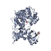 6ej8C  6ej9C 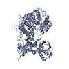 6ejaC 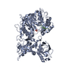 6ejbC 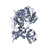 6ejcC 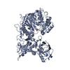 6ejdC  6ejeC  6foaC 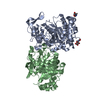 2gakS S: Starting model for refinement C: citing same article ( |
|---|---|
| Similar structure data |


-Links
 PDBj
PDBj


- Assembly
| Deposited unit | 
| ||||||||
|---|---|---|---|---|---|---|---|---|---|
| 1 |
| ||||||||
| Unit cell |
|
-Components
-Protein / Protein/peptide / Sugars , 3 types, 3 molecules AB
| #1: Protein | Mass: 86122.047 Da / Num. of mol.: 1 Source method: isolated from a genetically manipulated source Source: (gene. exp.) Homo sapiens (human) / Gene: XYLT1, XT1 / Plasmid: pCEP-Pu / Cell line (production host): HEK293F / Production host: Homo sapiens (human) / References: UniProt: Q86Y38, protein xylosyltransferase |
|---|---|
| #2: Protein/peptide | Mass: 1075.003 Da / Num. of mol.: 1 / Mutation: S215A, L220G, V221G / Source method: obtained synthetically / Source: (synth.) Homo sapiens (human) / References: UniProt: P02760 |
| #3: Sugar | ChemComp-NAG /  Type: D-saccharide, beta linking / Mass: 221.208 Da / Num. of mol.: 1 Type: D-saccharide, beta linking / Mass: 221.208 Da / Num. of mol.: 1Source method: isolated from a genetically manipulated source Formula: C8H15NO6 |
-Non-polymers , 6 types, 383 molecules 


| #4: Chemical | ChemComp-UDX /  Mass: 536.276 Da / Num. of mol.: 1 / Source method: obtained synthetically / Formula: C14H22N2O16P2 / Feature type: SUBJECT OF INVESTIGATION Mass: 536.276 Da / Num. of mol.: 1 / Source method: obtained synthetically / Formula: C14H22N2O16P2 / Feature type: SUBJECT OF INVESTIGATION | ||||||
|---|---|---|---|---|---|---|---|
| #5: Chemical | ChemComp-NA /  Mass: 22.990 Da / Num. of mol.: 1 / Source method: obtained synthetically / Formula: Na Mass: 22.990 Da / Num. of mol.: 1 / Source method: obtained synthetically / Formula: Na | ||||||
| #6: Chemical | ChemComp-PO4 /  Mass: 94.971 Da / Num. of mol.: 9 / Source method: obtained synthetically / Formula: PO4 Mass: 94.971 Da / Num. of mol.: 9 / Source method: obtained synthetically / Formula: PO4#7: Chemical | ChemComp-EDO /  Mass: 62.068 Da / Num. of mol.: 4 / Source method: obtained synthetically / Formula: C2H6O2 Mass: 62.068 Da / Num. of mol.: 4 / Source method: obtained synthetically / Formula: C2H6O2#8: Chemical |  Mass: 106.120 Da / Num. of mol.: 2 / Source method: obtained synthetically / Formula: C4H10O3 Mass: 106.120 Da / Num. of mol.: 2 / Source method: obtained synthetically / Formula: C4H10O3#9: Water | ChemComp-HOH / | Mass: 18.015 Da / Num. of mol.: 366 / Source method: isolated from a natural source / Formula: H2O |
-Details
| Has protein modification | Y |
|---|
-Experimental details
-Experiment
| Experiment | Method: X-RAY DIFFRACTION / Number of used crystals: 1 |
|---|
- Sample preparation
| Crystal | Density Matthews: 2.72 Å3/Da / Density % sol: 54.86 % |
|---|---|
| Crystal grow | Temperature: 293 K / Method: vapor diffusion, hanging drop / pH: 7.5 Details: 55-65% Morpheus precipitant mix 2 (PEG8000 and ethylene glycol), 0.1M Bicine/Tris buffer at pH 7.5 0.1 M of Morpheus NPS mix. |
-Data collection
| Diffraction | Mean temperature: 100 K | ||||||||||||||||||||||||
|---|---|---|---|---|---|---|---|---|---|---|---|---|---|---|---|---|---|---|---|---|---|---|---|---|---|
| Diffraction source | Source: SYNCHROTRON / Site: Diamond / Beamline: I04-1 / Wavelength: 0.9282 Å | ||||||||||||||||||||||||
| Detector | Type: DECTRIS PILATUS 6M-F / Detector: PIXEL / Date: Jan 21, 2017 | ||||||||||||||||||||||||
| Radiation | Protocol: SINGLE WAVELENGTH / Monochromatic (M) / Laue (L): M / Scattering type: x-ray | ||||||||||||||||||||||||
| Radiation wavelength | Wavelength: 0.9282 Å / Relative weight: 1 | ||||||||||||||||||||||||
| Reflection | Resolution: 2→86.89 Å / Num. obs: 61497 / % possible obs: 99.9 % / Redundancy: 6.5 % / Biso Wilson estimate: 30.33 Å2 / CC1/2: 0.996 / Rmerge(I) obs: 0.183 / Rpim(I) all: 0.077 / Rrim(I) all: 0.198 / Net I/σ(I): 9 / Num. measured all: 400825 / Scaling rejects: 0 | ||||||||||||||||||||||||
| Reflection shell | Diffraction-ID: 1
|
-Phasing
| Phasing | Method: molecular replacement |
|---|
- Processing
| Software |
| ||||||||||||||||||||||||||||||||||||||||||||||||||||||||||||||||||||||||||||||||||||||||||||||||||||||||||||||||||||||||||||||||||||||||||
|---|---|---|---|---|---|---|---|---|---|---|---|---|---|---|---|---|---|---|---|---|---|---|---|---|---|---|---|---|---|---|---|---|---|---|---|---|---|---|---|---|---|---|---|---|---|---|---|---|---|---|---|---|---|---|---|---|---|---|---|---|---|---|---|---|---|---|---|---|---|---|---|---|---|---|---|---|---|---|---|---|---|---|---|---|---|---|---|---|---|---|---|---|---|---|---|---|---|---|---|---|---|---|---|---|---|---|---|---|---|---|---|---|---|---|---|---|---|---|---|---|---|---|---|---|---|---|---|---|---|---|---|---|---|---|---|---|---|---|---|
| Refinement | Method to determine structure: MOLECULAR REPLACEMENT Starting model: 2GAK Resolution: 2→76.455 Å / SU ML: 0.28 / Cross valid method: FREE R-VALUE / σ(F): 1.34 / Phase error: 26.67
| ||||||||||||||||||||||||||||||||||||||||||||||||||||||||||||||||||||||||||||||||||||||||||||||||||||||||||||||||||||||||||||||||||||||||||
| Solvent computation | Shrinkage radii: 0.9 Å / VDW probe radii: 1.11 Å | ||||||||||||||||||||||||||||||||||||||||||||||||||||||||||||||||||||||||||||||||||||||||||||||||||||||||||||||||||||||||||||||||||||||||||
| Displacement parameters | Biso max: 141.04 Å2 / Biso mean: 45.9448 Å2 / Biso min: 19.28 Å2 | ||||||||||||||||||||||||||||||||||||||||||||||||||||||||||||||||||||||||||||||||||||||||||||||||||||||||||||||||||||||||||||||||||||||||||
| Refinement step | Cycle: final / Resolution: 2→76.455 Å
| ||||||||||||||||||||||||||||||||||||||||||||||||||||||||||||||||||||||||||||||||||||||||||||||||||||||||||||||||||||||||||||||||||||||||||
| Refine LS restraints |
| ||||||||||||||||||||||||||||||||||||||||||||||||||||||||||||||||||||||||||||||||||||||||||||||||||||||||||||||||||||||||||||||||||||||||||
| LS refinement shell | Refine-ID: X-RAY DIFFRACTION / Rfactor Rfree error: 0 / Total num. of bins used: 22 / % reflection obs: 100 %
| ||||||||||||||||||||||||||||||||||||||||||||||||||||||||||||||||||||||||||||||||||||||||||||||||||||||||||||||||||||||||||||||||||||||||||
| Refinement TLS params. | Method: refined / Refine-ID: X-RAY DIFFRACTION
| ||||||||||||||||||||||||||||||||||||||||||||||||||||||||||||||||||||||||||||||||||||||||||||||||||||||||||||||||||||||||||||||||||||||||||
| Refinement TLS group |
|
