 Movie
Movie Controller

[English] 日本語
 Yorodumi
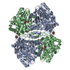
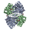
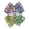
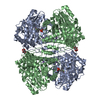
- PDB-6ecg: Citrobacter freundii tyrosine phenol-lyase F448A mutant complexed... -
+ Open data

- Basic information
| Entry | Database: PDB / ID: 6ecg | ||||||
|---|---|---|---|---|---|---|---|
| Title | Citrobacter freundii tyrosine phenol-lyase F448A mutant complexed with L-methionine | ||||||
 Components Components | (Tyrosine phenol- ...) x 2 | ||||||
 Keywords Keywords | LYASE / pyridoxal-5'-phosphate / aminotransferase fold | ||||||
| Function / homology |  Function and homology information Function and homology information | ||||||
| Biological species |  Citrobacter freundii (bacteria) Citrobacter freundii (bacteria) | ||||||
| Method |  X-RAY DIFFRACTION / SYNCHROTRON / MOLECULAR REPLACEMENT / Resolution: 2.27 Å X-RAY DIFFRACTION / SYNCHROTRON / MOLECULAR REPLACEMENT / Resolution: 2.27 Å | ||||||
 Authors Authors | Phillips, R.S. | ||||||
 Citation Citation | Journal: Biochemistry / Year: 2018 Title: Crystal Structures of Wild-Type and F448A Mutant Citrobacter freundii Tyrosine Phenol-Lyase Complexed with a Substrate and Inhibitors: Implications for the Reaction Mechanism. Authors: Phillips, R.S. / Craig, S. | ||||||
| History |
|
- Structure visualization
| Structure viewer | Molecule: MolmilJmol/JSmol |
|---|
- Downloads & links
-Download
| PDBx/mmCIF format | 6ecg.cif.gz | 430.1 KB | Display | PDBx/mmCIF format |
|---|---|---|---|---|
| PDB format | pdb6ecg.ent.gz | 352.5 KB | Display | PDB format |
| PDBx/mmJSON format | 6ecg.json.gz | Tree view | PDBx/mmJSON format | |
| Others |  Other downloads Other downloads |
-Validation report
| Arichive directory | https://data.pdbj.org/pub/pdb/validation_reports/ec/6ecgftp://data.pdbj.org/pub/pdb/validation_reports/ec/6ecg | HTTPS FTP |
|---|
-Related structure data
| Related structure data |  6durC 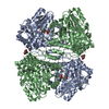 6dvxC  6dxvC 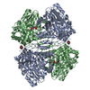 6dytC  6dz5C  2vlhS S: Starting model for refinement C: citing same article ( |
|---|---|
| Similar structure data |

-Links
 PDBj
PDBj- Assembly
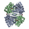
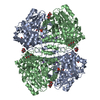
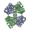
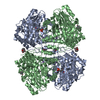
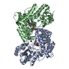
| Deposited unit | 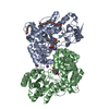
| ||||||||
|---|---|---|---|---|---|---|---|---|---|
| 1 | 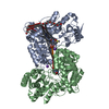
| ||||||||
| Unit cell |
|
-Components
-Tyrosine phenol- ... , 2 types, 2 molecules AB
| #1: Protein | Mass: 51301.461 Da / Num. of mol.: 1 / Mutation: F448A Source method: isolated from a genetically manipulated source Source: (gene. exp.) Citrobacter freundii (bacteria) / Gene: tpl / Plasmid: pLATE11-tpl / Production host: |
|---|---|
| #2: Protein | Mass: 51529.578 Da / Num. of mol.: 1 / Mutation: F448A Source method: isolated from a genetically manipulated source Source: (gene. exp.) Citrobacter freundii (bacteria) / Gene: tpl / Plasmid: pLATE11-tpl / Production host: |
-Non-polymers , 5 types, 654 molecules 
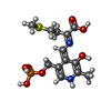


| #3: Chemical |  Mass: 39.098 Da / Num. of mol.: 2 / Source method: obtained synthetically / Formula: K Mass: 39.098 Da / Num. of mol.: 2 / Source method: obtained synthetically / Formula: K#4: Chemical | ChemComp-PM9 / ( |  Mass: 378.338 Da / Num. of mol.: 1 / Source method: obtained synthetically / Formula: C13H19N2O7PS Mass: 378.338 Da / Num. of mol.: 1 / Source method: obtained synthetically / Formula: C13H19N2O7PS#5: Chemical | ChemComp-PGE / |  Mass: 150.173 Da / Num. of mol.: 1 / Source method: obtained synthetically / Formula: C6H14O4 Mass: 150.173 Da / Num. of mol.: 1 / Source method: obtained synthetically / Formula: C6H14O4#6: Chemical | ChemComp-P33 / |  Mass: 326.383 Da / Num. of mol.: 1 / Source method: obtained synthetically / Formula: C14H30O8 / Comment: precipitant*YM Mass: 326.383 Da / Num. of mol.: 1 / Source method: obtained synthetically / Formula: C14H30O8 / Comment: precipitant*YM#7: Water | ChemComp-HOH / | Mass: 18.015 Da / Num. of mol.: 649 / Source method: isolated from a natural source / Formula: H2O |
|---|
-Experimental details
-Experiment
| Experiment | Method: X-RAY DIFFRACTION / Number of used crystals: 1 |
|---|
- Sample preparation
| Crystal | Density Matthews: 2.77 Å3/Da / Density % sol: 55.62 % |
|---|---|
| Crystal grow | Temperature: 293 K / Method: vapor diffusion, hanging drop / pH: 8 Details: 0.05 M triethanolamine-HCl, pH 8.0, 0.2 M KCl, 1 mM DTT, 0.5 mM pyridoxal-5'-phosphate |
-Data collection
| Diffraction | Mean temperature: 100 K |
|---|---|
| Diffraction source | Source: SYNCHROTRON / Site: APS  / Beamline: 22-ID / Wavelength: 1 Å / Beamline: 22-ID / Wavelength: 1 Å |
| Detector | Type: DECTRIS EIGER X 16M / Detector: PIXEL / Date: Dec 14, 2017 |
| Radiation | Protocol: SINGLE WAVELENGTH / Monochromatic (M) / Laue (L): M / Scattering type: x-ray |
| Radiation wavelength | Wavelength: 1 Å / Relative weight: 1 |
| Reflection | Resolution: 2.27→45.95 Å / Num. obs: 53790 / % possible obs: 99.92 % / Redundancy: 6 % / Biso Wilson estimate: 42.36 Å2 / CC1/2: 0.997 / Rmerge(I) obs: 0.1189 / Rpim(I) all: 0.05322 / Rrim(I) all: 0.1307 / Net I/σ(I): 7.75 |
| Reflection shell | Resolution: 2.27→2.351 Å / Redundancy: 6.2 % / Num. unique obs: 5280 / CC1/2: 0.239 / Rpim(I) all: 1.495 / Rrim(I) all: 3.751 / % possible all: 99.91 |
- Processing
| Software |
| ||||||||||||||||||||||||||||||||||||||||||||||||||||||||||||||||||||||||||||||||||||||||||||||||||||||||||||||||||||||||||||||||||||||||||||||||||||||||||||||||||||||||||||||||||||||||||||||||||||||||
|---|---|---|---|---|---|---|---|---|---|---|---|---|---|---|---|---|---|---|---|---|---|---|---|---|---|---|---|---|---|---|---|---|---|---|---|---|---|---|---|---|---|---|---|---|---|---|---|---|---|---|---|---|---|---|---|---|---|---|---|---|---|---|---|---|---|---|---|---|---|---|---|---|---|---|---|---|---|---|---|---|---|---|---|---|---|---|---|---|---|---|---|---|---|---|---|---|---|---|---|---|---|---|---|---|---|---|---|---|---|---|---|---|---|---|---|---|---|---|---|---|---|---|---|---|---|---|---|---|---|---|---|---|---|---|---|---|---|---|---|---|---|---|---|---|---|---|---|---|---|---|---|---|---|---|---|---|---|---|---|---|---|---|---|---|---|---|---|---|---|---|---|---|---|---|---|---|---|---|---|---|---|---|---|---|---|---|---|---|---|---|---|---|---|---|---|---|---|---|---|---|---|
| Refinement | Method to determine structure: MOLECULAR REPLACEMENT Starting model: 2VLH Resolution: 2.27→45.95 Å / SU ML: 0.32 / Cross valid method: THROUGHOUT / σ(F): 1.34 / Phase error: 24.24 / Stereochemistry target values: ML
| ||||||||||||||||||||||||||||||||||||||||||||||||||||||||||||||||||||||||||||||||||||||||||||||||||||||||||||||||||||||||||||||||||||||||||||||||||||||||||||||||||||||||||||||||||||||||||||||||||||||||
| Solvent computation | Shrinkage radii: 0.9 Å / VDW probe radii: 1.11 Å / Solvent model: FLAT BULK SOLVENT MODEL | ||||||||||||||||||||||||||||||||||||||||||||||||||||||||||||||||||||||||||||||||||||||||||||||||||||||||||||||||||||||||||||||||||||||||||||||||||||||||||||||||||||||||||||||||||||||||||||||||||||||||
| Displacement parameters | Biso mean: 57.24 Å2 | ||||||||||||||||||||||||||||||||||||||||||||||||||||||||||||||||||||||||||||||||||||||||||||||||||||||||||||||||||||||||||||||||||||||||||||||||||||||||||||||||||||||||||||||||||||||||||||||||||||||||
| Refinement step | Cycle: LAST / Resolution: 2.27→45.95 Å
| ||||||||||||||||||||||||||||||||||||||||||||||||||||||||||||||||||||||||||||||||||||||||||||||||||||||||||||||||||||||||||||||||||||||||||||||||||||||||||||||||||||||||||||||||||||||||||||||||||||||||
| LS refinement shell |
| ||||||||||||||||||||||||||||||||||||||||||||||||||||||||||||||||||||||||||||||||||||||||||||||||||||||||||||||||||||||||||||||||||||||||||||||||||||||||||||||||||||||||||||||||||||||||||||||||||||||||
| Refinement TLS params. | Method: refined / Refine-ID: X-RAY DIFFRACTION
| ||||||||||||||||||||||||||||||||||||||||||||||||||||||||||||||||||||||||||||||||||||||||||||||||||||||||||||||||||||||||||||||||||||||||||||||||||||||||||||||||||||||||||||||||||||||||||||||||||||||||
| Refinement TLS group |
|
